What treatment


Highly Recommended Dentistry in Turkey Articles
Explore Our Trending Dentistry in Turkey Articles
Trusted Medical Tourism Platform Since 2007

5 Turkish Cities for Dental Tourism Compared: Istanbul vs Antalya vs Izmir for European Patients

Overview
This comparison evaluates five major Turkish cities for dental tourism: Istanbul’s vast clinic choice, Antalya’s beach recovery, Izmir’s coastal value, Ankara’s academic centers, and Bodrum’s exclusive retreats, helping European patients match dental excellence with their preferred holiday style....
Read more details.jpeg)

Full Mouth Zirconium Crown Makeover in Turkey: 7 Steps UK Patients Follow From Booking to Results
Overview
This article maps the seven step journey UK patients undertake for a full mouth zirconium crown makeover in Turkey. It details the initial video consult, digital smile design, temporary wear, final monolithic zirconia preparation, bonding, occlusion check, and the sparkling final reveal....
Read more details
E-max vs Zirconium Veneers in Turkey: 7 Facts UK Cosmetic Patients Need to Decide
Overview
Seven factual comparisons help UK cosmetic patients decide between E-max and zirconium veneers in Turkey. This article contrasts aesthetic translucency for anterior teeth vs. zirconium’s posterior strength, minimal prep techniques, and laboratory cementation protocols for long lasting results....
Read more details
10 Reasons German Patients Are Saving Up to €24,000 on Hollywood Smile Veneers in Turkey
Overview
Ten compelling reasons lead German patients to save up to €24,000 on Hollywood smile veneers in Turkey. This overview highlights premium E-max and laminate materials, digital smile design, German speaking patient coordinators, and comprehensive aftercare that ensures a red carpet ready smile....
Read more details
All-on-4 vs All-on-6 in Turkey: 5 Key Differences UK Patients Must Know Before Booking
Overview
Before booking full arch restoration in Turkey, UK patients must compare five key differences between All on 4 and All on 6. This article examines implant number impact on load distribution, bone grafting necessity, overall fee, and which option delivers the most durable, aesthetic outcome for their case....
Read more detailsDiscover your treatment options with a free, no-obligation quote!
Get your quote now!
7 Reasons UK Patients Choose Turkey for Single Dental Implants Over NHS Waiting Lists
Overview
Seven key reasons lead UK patients to Turkey for single dental implants instead of queuing on the NHS. Immediate implant placement, digital smile design, premium implant brands, and transparent all inclusive pricing provide a faster, more predictable tooth replacement journey....
Read more details
Same-Day Teeth in Turkey vs Greece - Immediate Load Implants, Holiday Experiences, and Clinic Guarantees for UK Tourists
Overview
Choosing between Turkey and Greece for same-day teeth involves balancing exceptional cost savings, immediate load implant expertise, and appealing holiday environments. This comprehensive guide compares clinic guarantees, treatment timelines, and exact pricing to help UK tourists make an informed, confident decision for their full mouth restoration....
Read more details
Dental Bridges in Turkey vs Czech Republic - Material Quality, Lab Turnaround Times, and Costs for Austrian Patients
Overview
For Austrian patients facing high domestic dental costs, traveling abroad for restorative dentistry has become a highly practical solution. This guide provides a direct comparison of dental bridges in Turkey vs Czech Republic, two of the leading destinations for high-quality, affordable dental care. We will evaluate critical factors including exact procedure costs, dental laboratory turnaround times, and the quality of materials used, helping you make a confident, well-informed decision for your oral health....
Read more details
Cosmetic Dentistry in Turkey vs Dubai - Luxury Clinic Experiences vs Budget Affordability for UAE Expats
Overview
For United Arab Emirates expatriates seeking a flawless smile, the decision often narrows down to two premier destinations: remaining local for ultra-luxury dental care in Dubai or traveling abroad for highly affordable, world-class dental tourism in Turkey. This comprehensive comparison analyzes costs, clinic experiences, material quality, and logistical considerations to help you choose the ideal destination for your cosmetic dentistry journey....
Read more detailsDiscover your treatment options with a free, no-obligation quote!
Get your quote now!
Straumann Implants in Turkey vs Germany - Aftercare Risks, 70% Price Drops, and Brand Guarantees for German Citizens
Overview
While Germany offers unparalleled localized aftercare and stringent regulatory protection for dental implant procedures, Turkey provides identical premium Straumann implants at up to a 70% price reduction. The ultimate decision for German citizens hinges on balancing upfront cost savings against travel logistics, cross-border aftercare risks, and leveraging global brand guarantees....
Read more details
Emax Veneers in Turkey vs Romania - VIP Packages, Hotel Inclusions, and Quality Comparisons for UK Patients
Overview
Deciding between Emax veneers in Turkey and Emax veneers in Romania requires evaluating VIP packages, hotel inclusions, overall dental quality, and total procedure costs. Both destinations offer exceptional dental tourism opportunities for UK patients seeking premium smile makeovers at a fraction of domestic prices, but they differ significantly in patient experience, European Union regulatory frameworks, and the style of all-inclusive hospitality provided....
Read more details
Dental Implants in Turkey vs Spain - Recovery Environments, Hidden Clinic Fees, and Quality Standards for British Retirees
Overview
For British retirees facing exorbitant private dental fees and extensive NHS wait times, seeking restorative dentistry abroad has become a primary solution. Deciding between dental implants in Turkey vs Spain is a critical choice that goes far beyond simple price tags. Both destinations offer remarkable savings and top-tier clinical expertise, but they cater to different patient needs regarding travel logistics, post-operative environments, and regulatory frameworks....
Read more details.png)
Smile Makeovers in Turkey vs Croatia - All-Inclusive Packages, Zirconia Brands, and Safety for Irish Patients
Overview
For Irish patients seeking affordable, high-quality dental restoration, choosing between a Smile Makeover in Turkey and a Smile Makeover in Croatia is a major decision. Both nations offer exceptional dental tourism opportunities, but they cater to different patient preferences regarding all-inclusive packages, EU safety regulations, and clinic environments. This comprehensive comparison will help you navigate the exact pricing, global zirconia brands, and safety protocols necessary to make the best choice for your dental health and travel comfort....
Read more details
Zirconia Crowns in Turkey vs Poland - Price vs EU Standards: A Complete Guide for German & Scandinavian Dental Tourists
Overview
For German and Scandinavian dental tourists, choosing between Zirconia crowns in Turkey and Poland comes down to prioritizing either maximum cost savings with luxury VIP experiences or strict adherence to European Union medical standards. While Turkey offers unbeatable all-inclusive packages and rapid turnaround times, Poland provides legally binding EU dental warranties, closer geographical proximity, and highly regulated, conservative dental treatments....
Read more details
Porcelain Veneers in Turkey vs UK Private Care - NHS Wait Times, 'Turkey Teeth' Risks, and Real Savings for UK Residents
Overview
Navigating the decision between UK private dental care and seeking porcelain veneers in Turkey requires balancing significant cost savings against potential risks. This comprehensive guide compares procedural standards, the reality of NHS wait times, the infamous "Turkey teeth" risks, and actionable insights to help UK residents make an informed, safe decision for their smile makeover....
Read more details
All-on-4 Dental Implants in Turkey vs Hungary - Cost Breakdowns, EU Warranties, and Clinic Quality for British Patients
Overview
For British patients facing skyrocketing private dental fees and extensive NHS wait times, traveling abroad for full mouth restorations has become a highly attractive, practical solution. Choosing the right destination for a procedure as significant as All-on-4 dental implants is a monumental decision, requiring a careful evaluation of treatment costs, clinic quality, and post-operative protections. Today, two destinations dominate the European dental tourism landscape: Turkey and Hungary. This comprehensive comparison will break down the nuances of pursuing All-on-4 dental implants in Turkey vs Hungary, providing actionable insights into cost breakdowns, EU warranties, and overall clinic quality to help UK patients navigate their dental restoration journey with confidence....
Read more details
Why Izmir is Top Choice 2026 for Eco-Friendly and Bio-Compatible Dental Implants?
Overview
The 'Sustainability' Smile: Why Izmir is 2026’s Top Choice for Eco-Friendly, Bio-Compatible Dental Implants,"Izmir is 2026’s top choice for the sustainability smile—eco-friendly, metal-free zirconia implants that are fully biocompatible, appealing to health-conscious patients wanting a green, holistic dental solution."...
Read more details
One-Hour Excellence: How Istanbul’s 2026 In-House Labs are Eliminating the Need for Temporary Dental Crowns
Overview
One-Hour Excellence: How Istanbul’s 2026 In-House Labs are Eliminating the Need for Temporary Dental Crowns,"Istanbul’s in-house labs achieve one-hour excellence—designing, milling, and placing permanent ceramic crowns in a single visit, completely removing the inconvenience of temporary crowns for international patients."...
Read more details.jpg)
Why is Istanbul Dominating the 2026 Market for Full-Mouth Rehabilitations?
Overview
The Global Hub for Fixed Arches: Why Istanbul is Dominating the 2026 Market for Full-Mouth Rehabilitations,"Istanbul dominates 2026 as the global hub for fixed arches—full-mouth implant rehabilitations (All-on-4/6) delivered with advanced technology, master ceramists, and all-inclusive packages at competitive prices."...
Read more details
Why 2026 Antalya Trends Favor Translucent E-Max Over Opaque Dental Crowns?
Overview
Zirconia Excellence: Why 2026 Antalya Trends Favor Translucent E-Max Over Opaque Dental Crowns,"2026 Antalya trends favor translucent E-Max crowns over opaque alternatives—zirconia excellence that mimics natural enamel, delivering strength, beauty, and the most lifelike appearance for dental restorations."...
Read more detailsShare with AI
Share this page with AI assistants to get summaries and insights
Table of Contents
- 5 Turkish Cities for Dental Tourism Compared: Istanbul vs Antalya vs Izmir for European Patients
- Full Mouth Zirconium Crown Makeover in Turkey: 7 Steps UK Patients Follow From Booking to Results
- E-max vs Zirconium Veneers in Turkey: 7 Facts UK Cosmetic Patients Need to Decide
- 10 Reasons German Patients Are Saving Up to €24,000 on Hollywood Smile Veneers in Turkey
- All-on-4 vs All-on-6 in Turkey: 5 Key Differences UK Patients Must Know Before Booking
Explore : Stories, Testimonials, and Procedures
Watch in-depth videos covering everything from success stories to detailed procedure explanations. Learn what to expect and hear from real patients.
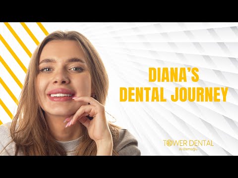
Transforming Your Smile with Dental Veneers in Istanbul, Turkey
Dentistry
Tower Dental Clinic
Istanbul, Turkey
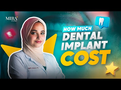
How Much Do Dental Implants Cost in Turkey? Everything You Need to Know! DR.Lubaba Alhaj from MIRA clinic
Dentistry
MIRA Clinic - Dental Treatments in Istanbul, Turkey
Istanbul, Turkey

Comprehensive Guide to the Full Mouth Dental Implants Procedure in Istanbul, Turkey
Dentistry
YEG Clinic - Best Dental Clinic Istanbul
Istanbul, Turkey
Complete Guide to Achieving a Flawless Smile Makeover with Porcelain Veneers in Istanbul, Turkey
Dentistry
YEG Clinic - Best Dental Clinic Istanbul
Istanbul, Turkey
Unlock the Best Packages Just for You
Discover the ultimate packages designed to meet your health and wellness goals. Choose the perfect fit for your journey.

Price starting from $3,800
Hollywood Smile Package in Istanbul, Turkey by YEG Clinic
YEG Clinic - Best Dental Clinic Istanbul
- Dentistry
Istanbul, Turkey

Price starting from $7,500
Dental Implants Package in Istanbul, Turkey at Medical Park Dental Clinics
Medical Park Hospitals Group in Istanbul, Turkey
- Dentistry
Istanbul, Turkey
.png)
Price starting from $1,000
Cosmetic Dentistry in Turkey by Phinova Clinics - Dental Clinic Izmir
Phinova Clinics - Dental Clinic Izmir
- Dentistry
Izmir, Turkey
.png)
Price starting from $1,000
Budget-Friendly Cosmetic Dentistry Package in Turkey by OGN Dental Studio
OGN Dental Studio - Dental Center Antalya Turkey
- Dentistry
Antalya, Turkey
Transformative Experiences: Dive into Procedure Insights
Discover the power of through compelling patient experiences. Uncover detailed insights into procedures, recovery journeys, and life-changing results.
.png)
.png)
.png)
.png)