
Addressing Complex Tumors: Tumor Removal Surgery in Saudi Arabia
Complex surgical interventions for rare and extensive tumors that extend through major blood vessels into the heart require advanced surgical expertise, meticulous planning, and seamless coordination among multiple medical specialties. Discover how multidisciplinary teams for Tumor Removal Surgery in Saudi Arabia at leading medical centers, King Faisal Specialist Hospital and Research Centre (KFSHRC), approach challenging tumor removal procedures, combining specialized surgical techniques, cardiovascular intervention, and vascular reconstruction to address complex anatomical involvement, restore blood vessel integrity, and support patient recovery through comprehensive perioperative care.

Quick Summary: Single-Stage Multidisciplinary Surgical Case
- Patient Profile: A woman in her forties presenting with an extensive intravenous tumor.
- Medical Diagnosis: Rare intravenous leiomyomatosis originating in the uterus and advancing into the cardiovascular system.
- Extent of Tumorous Invasion: Tumor extended through the inferior vena cava, right atrium of the heart, hepatic veins, and major pelvic blood vessels.
- Surgical Complexity: Over 16 hours of continuous, single-stage operation involving gynecologic oncologists, vascular surgeons, and cardiac surgeons.
- Surgical Execution: Complete tumor resection, hysterectomy, and reconstruction of affected blood vessels and the heart wall.
- Clinical Outcome: Successfully eliminated the need for staged surgeries, significantly minimizing operative complications and supporting faster recovery.
Overview of Life-Saving Multidisciplinary Tumor Removal Surgery in Saudi Arabia
Severe intravascular neoplasms that extend from the pelvis directly into the thoracic cavity present some of the most critical challenges in modern surgical oncology. Executing a life-saving multidisciplinary Tumor Removal Surgery in Saudi Arabia requires extreme surgical precision, advanced cardiopulmonary bypass infrastructure, and real-time intraoperative coordination across multiple surgical specialties.
When tumors invade primary blood vessels and reach cardiac chambers, staged operations often carry heightened risks of pulmonary embolism, severe hemorrhage, or tumor fragment detachment. Performing a comprehensive single-stage resection offers a decisive approach that addresses both the primary origin and intravascular extensions simultaneously.
Understanding the Extensive Tumor Reaching the Heart
Intravenous leiomyomatosis is an exceptionally rare, histologically benign yet biologically aggressive neoplasm that arises from uterine smooth muscle tissue. Rather than invading local tissue directly, the tumor grows along the lumen of uterine and iliac veins, migrating into the body's primary venous pathways.
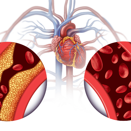
In severe clinical presentations, performing Intravenous Leiomyomatosis Surgery in Saudi Arabia becomes mandatory when the neoplastic mass extends upwards through the inferior vena cava (IVC), reaches the right atrium of the heart, and occludes hepatic veins. Without immediate surgical intervention, complete intravascular occlusion can lead to right heart failure or sudden fatal cardiac events.
Primary Pelvic Origin
Originates within uterine smooth muscle tissue, infiltrating uterine and internal iliac veins.
Vascular Migration
Tracks superiorly through the inferior vena cava, impairing venous return and hepatic clearance.
Intracardiac Extension
Reaches the right atrium, creating severe hemodynamics obstruction within cardiac chambers.
Patient Presentation and Diagnostic Evaluation
A woman in her forties presented with progressive physical exhaustion, lower extremity edema, and exertional shortness of breath. Because symptoms of intravascular tumors often mimic primary cardiac conditions or deep vein thrombosis, thorough clinical evaluation is essential to establish the true extent of disease.
Initial physical examination and blood panels led physicians to investigate central venous blood flow. Diagnostic assessments revealed a continuous fibrous tumor tracking from the pelvic floor straight into the right side of the heart, requiring immediate referral for specialized Tumor Surgery in Saudi Arabia.
Advanced Imaging and Tumor Assessment
Mapping an extensive intravascular tumor requires multi-modality diagnostic imaging. Radiologists utilize contrast-enhanced Computed Tomography (CT), Magnetic Resonance Imaging (MRI), and Echocardiography to evaluate tumor adherence, vascular wall infiltration, and cardiac mobility.
| Imaging Modality | Clinical Assessment Purpose |
|---|---|
| 3D Contrast CT Venography | Maps total tumor length from pelvic veins through the inferior vena cava to the heart. |
| Cardiac MRI | Differentiates tumor tissue from blood clots and checks for direct myocardial invasion. |
| Transesophageal Echocardiogram (TEE) | Provides real-time visualization of right atrial motion, tricuspid valve interaction, and cardiac obstruction. |
Global Trends in Complex Multidisciplinary Surgeries
Data published by global health monitoring organizations highlight the growing volume of complex organ-reconstruction and multi-specialty tumor procedures conducted at tertiary research centers worldwide.
Worldwide Complex Multidisciplinary Surgical Procedures (WHO Estimate)
Source: World Health Organization (WHO) Surgical Care Global Report. Values reflect estimated worldwide complex multi-specialty cases.
Multidisciplinary Medical Team and Treatment Planning
Assembling a collaborative multi-specialty surgical team was critical to establishing a safe surgical roadmap. At KFSHRC Saudi Arabia, tumor boards consisting of cardiac surgeons, vascular surgeons, gynecologic oncologists, hepatic specialists, cardiac anesthesiologists, and perfusionists convened to plan the complex intervention.
The team selected a single-stage operative plan rather than divided procedures. This strategy reduced the cumulative burden of anesthesia, eliminated the risk of intervening tumor embolization, and enabled simultaneous vascular and pelvic control.
Challenges of Removing a Tumor Near the Heart
Resecting intravascular tumor extensions within cardiac structures presents serious surgical challenges. Mobilizing a mass that spans from the lower abdomen to the chest involves distinct intraoperative hazards:
Severe Hemorrhage Risk
Opening major venous channels like the inferior vena cava demands precise blood conservation and bypass control.
Tumor Embolization
Manipulation of the mass can cause tumor fragments to break off, risking fatal pulmonary arterial blockage.
Hemodynamic Instability
Clamping central veins abruptly drops blood return to the heart, requiring continuous pharmacological and pump support.
Surgical Approach to Extensive Tumor Removal
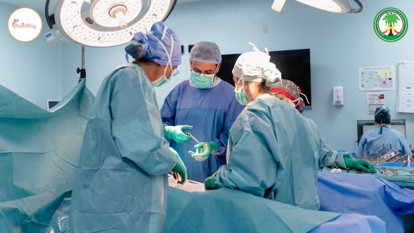
The operation required a continuous surgical duration exceeding 16 hours. Operating teams gained simultaneous access to the chest, abdomen, and pelvis through combined sternotomy and laparotomy incisions, allowing full control over the central circulatory system.
While gynecologic oncologists resected the primary uterine mass within the pelvis, cardiac and vascular teams established cardiopulmonary bypass. The tumor was extracted en bloc from the right atrium and inferior vena cava, followed by comprehensive hysterectomy to eliminate the neoplastic root.
Specialized Surgical Techniques and Medical Expertise
Removing extensive intravascular tumors can involve delicate blood vessels and cardiac tissues, requiring careful surgical planning and coordination across multiple specialties. After tumor extraction, cardiac and vascular reconstruction may be necessary to repair affected structures and support the restoration of normal blood flow.
Key Surgical Procedures
Tumor Extraction and Cardiac Repair
Following tumor removal, Cardiac Reconstruction Surgery in Saudi Arabia may involve repairing affected cardiac structures, including the right atrium, and reconstructing damaged sections of the vena cava using suitable surgical techniques and patch materials.
Vascular Reconstruction
Specialized surgical repair may address affected venous structures, including the hepatic and iliac venous pathways, depending on the tumor's extent and the anatomy involved.
Restoring Blood Flow and Monitoring Recovery
After reconstruction, the surgical team evaluates blood vessel patency, circulation, and cardiac function. Postoperative monitoring helps identify complications such as thrombosis and guides follow-up care.
Clinical consideration: The surgical approach depends on the tumor's location, extent of vascular involvement, and the patient's condition. Specific techniques and outcomes should be confirmed against the treating hospital's published case details.
Managing Cardiovascular Risks During Surgery
To protect brain and organ function during central vascular reconstruction, cardiac anesthesiologists utilized deep hypothermic circulatory arrest (DHCA) and targeted hypothermia. Lowering body temperature reduces cellular oxygen demand while surgeons safely resect tumor tissue from blood vessel walls.
Intraoperative Protocol: Advanced cell-saver blood conservation systems, continuous transesophageal echocardiography monitoring, and heparinization management were maintained throughout the 16-hour procedure to protect vital organ perfusion.
Postoperative Care, Intensive Monitoring, and Recovery
Following successful resection, the patient was transferred to the Cardiovascular Intensive Care Unit (CVICU). Specialized critical care teams provided continuous hemodynamic monitoring, mechanical ventilation support, anticoagulant management, and early mobilization protocols.
By consolidating the treatment into a single comprehensive procedure, the patient avoided the cumulative risks of multiple operations. Postoperative diagnostic imaging confirmed total tumor clearance, unhindered venous blood flow, and robust recovery of normal heart function.
The Role of Advanced Cancer Surgery at King Faisal Specialist Hospital and Research Centre
The successful management of this complex case highlights the tertiary surgical capabilities available at King Faisal Specialist Hospital and Research Centre. As a primary specialized research center in the Middle East, the institution integrates advanced clinical research, hybrid operating suites, and expert multi-specialty medical teams.
For international and regional patients seeking specialized treatment, accessing advanced care at KFSHRC involves comprehensive pre-arrival medical record reviews, teleconsultations, and structured travel coordination.
Indicative Financial Budget Components (USD)
Complex surgical care expenses depend on individual case requirements, operative hours, intensive care duration, and specialized implant materials. All financial estimates are structured in USD:
| Cost Component | Clinical Coverage | Financial Considerations (USD) |
|---|---|---|
| Preoperative Evaluation | 3D CT venography, cardiac MRI, transesophageal echo, blood panels | Itemized diagnostic testing rates |
| Surgical & Anesthesia Fees | Multi-specialty surgical teams (Gynecology, Vascular, Cardiac) | Based on operative time exceeding 16 hours |
| Cardiopulmonary Bypass & DHCA | Perfusionist support, hypothermia management, cell-saver systems | Specialized cardiac technology utilization |
| Cardiothoracic ICU Care | 1-on-1 intensive nursing, ventilator support, arterial line monitoring | Daily CVICU room and monitoring fees |
| Vascular Reconstruction Materials | Biological patches, synthetic grafts, cardiac reconstruction supplies | Surgical material itemization |
Frequently Asked Questions
What is intravenous leiomyomatosis?
Intravenous leiomyomatosis is an uncommon condition where benign uterine smooth muscle tissue grows into pelvic veins and extends upwards into central blood vessels, sometimes reaching the heart.
Why is a single-stage operation preferred for extensive tumors?
A single-stage operation allows surgeons to completely extract the tumor from both pelvic and cardiac structures in one procedure, eliminating the danger of tumor embolization between staged surgeries.
Which surgical specialists are required for this procedure?
The multidisciplinary team typically includes cardiac surgeons, vascular surgeons, gynecologic oncologists, cardiac anesthesiologists, perfusionists, and critical care specialists.
How long does complex multidisciplinary tumor removal take?
Due to the necessity of vascular isolation, cardiac bypass, and reconstruction, these complex surgical procedures can exceed 16 hours of continuous operating time.
Is cardiopulmonary bypass necessary during tumor extraction from the heart?
Yes, when tumors enter the right atrium, heart-lung machine bypass is required so surgeons can safely open cardiac chambers and remove the mass under direct visualization.
What is cardiac reconstruction surgery?
Cardiac reconstruction involves repairing or replacing damaged heart wall tissues, valves, or major attached vessels using biological or synthetic patches after tumor resection.
What diagnostic tests confirm tumor extension into blood vessels?
Contrast-enhanced CT venography, cardiac MRI, and transesophageal echocardiography (TEE) are the primary imaging tools used to define tumor boundaries.
What are the main risks associated with intravascular tumor surgery?
Major risks include severe intraoperative hemorrhage, tumor fragmentation embolization, venous thrombosis, systemic infection, and temporary organ dysfunction.
How long is the ICU recovery period after 16-hour tumor surgery?
Patients typically remain in the CVICU for several days under continuous monitoring before transitioning to standard inpatient surgical wards for progressive rehabilitation.
How can foreign patients request surgical consultation in Saudi Arabia?
International patients can submit detailed medical records, diagnostic imaging, and pathology reports through official referral channels for multi-specialty board review.
Consult King Faisal Specialist Hospital & Research Centre
Inquire about specialized multidisciplinary tumor evaluation, advanced cardiovascular surgery, and tertiary medical reviews at King Faisal Specialist Hospital and Research Centre in Riyadh.
Request Surgical InformationMedical Disclaimer: The information provided in this document is intended for educational and medical informational purposes only and does not constitute formal medical advice, clinical diagnosis, or treatment recommendations. Complex medical decisions regarding extensive tumor removal or cardiovascular surgery must always be made in direct consultation with qualified medical professionals and surgical specialists.
References:
- WHO: Cancer Diagnosis, Treatment, and Care
- CDC: Cancer Information and Treatment Resources
- Saudi Ministry of Health: Patient Education and Health Information
- PubMed: Research on Intravenous Leiomyomatosis and Cardiac Involvement
- American Heart Association: Cardiovascular Conditions and Treatment
- American Thoracic Society Journals: Cardiopulmonary Research


















Share this listing