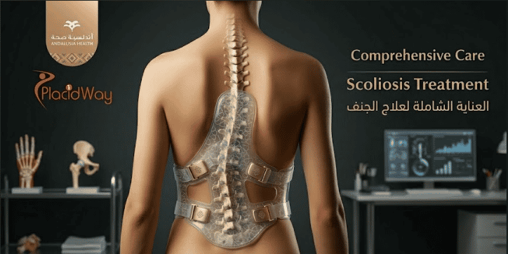
Orthopedic Technology Innovations: Revolutionizing Joint and Spine Surgery
The landscape of musculoskeletal medicine is undergoing a profound transformation driven by rapid orthopedic technology innovations. For decades, patients requiring joint replacement or complex spinal interventions faced long recovery times, significant surgical trauma, and the limitations of standardized medical implants.
Today, the integration of advanced robotics, artificial intelligence, and biological engineering has fundamentally rewritten the rules of surgical intervention. Watch as we explore how these cutting-edge digital and mechanical advancements are providing unprecedented precision, personalizing patient care, and dramatically accelerating post-operative rehabilitation.
Video Chapters & Quick Navigation
The convergence of engineering and medicine has pushed the boundaries of what is possible in treating musculoskeletal conditions. Patients suffering from severe osteoarthritis, traumatic sports injuries, or degenerative disc disease are no longer restricted to traditional, highly invasive procedures. Instead, they are stepping into an era defined by extreme precision, personalized anatomical modeling, and biological joint preservation.
Understanding these advancements is crucial for anyone facing joint replacement or spinal surgery. By exploring the mechanisms behind these high-tech interventions, patients can make highly informed decisions regarding their treatment pathways. The paradigm has shifted from simply replacing damaged bones to perfectly replicating native joint kinematics and restoring uncompromised mobility.
Robotic-Assisted Joint Replacement Surgery: Sub-Millimeter Precision
As detailed at in the video, robotic-assisted joint replacement surgery represents one of the most significant leaps forward in modern operative care. While traditional manual instrumentation relies heavily on the surgeon’s visual estimation and tactile feedback, robotic systems introduce an unparalleled level of mathematical accuracy to the operating table.
These sophisticated robotic platforms utilize advanced 3D anatomical mapping to create a virtual model of the patient's specific joint structure. Surgeons use this digital twin to pre-plan the exact size, orientation, and alignment of the prosthetic implant before a single incision is ever made. This ensures that the final placement perfectly mimics the patient's natural joint mechanics.
Haptic Boundary Technology and Soft Tissue Preservation
One of the most remarkable features of modern robotic orthopedic systems is the implementation of haptic boundary technology. Once the surgical plan is locked into the computer, the robotic arm establishes invisible virtual boundaries around the target bone. If the surgeon attempts to move the cutting tool outside of these safe zones, the robotic arm will automatically power down or resist the movement.
This critical safety mechanism ensures absolute protection for surrounding soft tissues, including delicate ligaments, nerves, and blood vessels. Because the surgical trauma is strictly confined to the diseased bone, patients experience drastically reduced post-operative pain. Furthermore, the preservation of key ligaments allows for a much more natural-feeling joint and a highly accelerated physical therapy timeline.
3D-Printed Orthopedic Implants: The End of "One Size Fits All"
Historically, orthopedic surgeons had to select implants from a predetermined inventory of standard sizes, often requiring them to alter the patient's bone to fit the available prosthesis. As highlighted at , the advent of 3D-printed orthopedic implants has completely reversed this paradigm. Now, the implant is specifically manufactured to fit the unique geometry of the patient's bone.
Using high-resolution CT scans, engineers can generate a precise computer-aided design (CAD) file of the patient's damaged joint. Additive manufacturing processes, specifically titanium powder bed fusion, are then used to build the custom implant layer by microscopic layer. This level of customization is particularly revolutionary for complex revision surgeries or massive bone loss due to orthopedic oncology.
Trabecular Metal and Enhanced Osseointegration
The true brilliance of 3D printing in orthopedics lies in the ability to manipulate the microscopic structure of the implant's surface. Traditional milled titanium implants have relatively uniform, roughened surfaces. Conversely, 3D printers can construct highly complex, porous structures that exactly mimic the trabecular architecture of natural human cancellous bone.
This highly porous biomaterial creates an ideal environment for osseointegration. The patient's natural bone tissue actively grows into the pores of the titanium implant, creating a biological fixation that is vastly superior to traditional bone cement. This living connection between bone and metal ensures the long-term survivorship of the implant, significantly reducing the likelihood of future implant loosening.
Artificial Intelligence in Orthopedic Diagnostics and Planning
Artificial intelligence in orthopedic diagnostics is fundamentally changing how rapidly and accurately conditions are diagnosed. As noted at , machine learning algorithms are now capable of analyzing thousands of radiographic images in mere seconds. These AI networks have been trained on vast datasets to detect microscopic fractures, subtle cartilage degradation, and early-stage joint space narrowing that might escape the human eye.
Beyond simple diagnostics, AI is playing a massive role in predictive surgical analytics. By inputting a patient's specific biometric data—such as age, BMI, bone density, and activity level—predictive AI models can forecast the likely outcomes of different surgical interventions. This allows the surgical team to choose the specific implant material and surgical approach that offers the highest statistical probability of long-term success.
Automated Pre-Operative Templating
Traditional pre-operative templating required the surgeon to manually overlay digital implant templates onto x-rays to estimate sizes. Today, AI-driven software completely automates this process. The system automatically segments the 3D bone models, instantly calculates optimal anatomical alignment, and suggests the exact implant size required.
This automation dramatically reduces pre-operative planning time while simultaneously increasing the accuracy of the selected components. When surgeons enter the operating room, they already have a mathematically optimized, AI-verified roadmap for the entire procedure. This streamlining reduces the total time under anesthesia, further improving overall patient safety.
Smart Implants and Wearable Technology for Rehabilitation
The post-operative recovery phase has traditionally been a black box for orthopedic surgeons. Once the patient left the hospital, clinicians had to rely on subjective patient reporting to monitor progress. As discussed at , the introduction of smart knee replacement technology and advanced wearables has illuminated this critical recovery period.
FDA-approved smart implants now feature miniaturized sensor stems embedded directly into the tibial extension of the joint replacement. These sensors contain advanced inertial measurement units (IMUs) that capture precise kinematic data with every step the patient takes. This revolutionary internal technology requires no charging, as the battery is engineered to outlast the typical lifespan of the monitoring period.
Remote Patient Monitoring and Data-Driven Therapy
The data collected by these smart implants—including range of motion, stride length, walking speed, and step count—is transmitted securely via a base station to a cloud-based clinical portal. Physical therapists and orthopedic surgeons can now review objective, real-time biomechanical data from their remote dashboards.
If a patient's recovery trajectory deviates from expected norms, the clinical team is immediately alerted. They can intervene early to adjust physical therapy protocols, address potential stiffness, or investigate emerging complications before they require readmission. This constant stream of telemetry ensures that rehabilitation is highly personalized and rigorously data-driven.
Orthobiologics and Regenerative Medicine in Joint Repair
While metal and plastic implants have excellent track records, the holy grail of orthopedics is biological joint preservation. Highlighted at , orthobiologics represent a shift away from replacing damaged tissues and moving toward regenerating them using the body's own healing mechanisms.
Advanced biologic treatments for joint repair utilize highly concentrated biological agents to stimulate cellular growth and tissue repair. These therapies are particularly effective in treating early-stage osteoarthritis, chronic tendinopathies, and focal cartilage defects that are not yet severe enough to warrant a total joint replacement.
Platelet-Rich Plasma and Mesenchymal Stem Cells
The most common applications of orthobiologics involve Platelet-Rich Plasma (PRP) and Mesenchymal Stem Cell (MSC) therapies. By drawing a small amount of the patient's blood or bone marrow, clinicians use specialized centrifuges to isolate powerful growth factors and unspecialized stem cells. These concentrated biological solutions are then precisely injected into the damaged joint space under ultrasound guidance.
Once introduced to the avascular zones of the joint, such as the meniscus or articular cartilage, these growth factors initiate a profound anti-inflammatory response. They actively recruit local repair cells and provide the biological scaffolding necessary to synthesize new, healthy tissue. This regenerative approach helps delay or even entirely prevent the need for invasive metallic joint replacements.
Minimally Invasive Surgical Techniques in Spine and Joint Care
The evolution of surgical instrumentation has paved the way for ultra-minimally invasive surgical techniques. Mentioned at , modern arthroscopic and endoscopic procedures have drastically reduced the collateral tissue damage associated with traditional open surgeries. The goal is to achieve maximal surgical correction through minimal anatomical disruption.
In the realm of spinal care, minimally invasive spine surgery techniques utilize progressive tubular retractors. Instead of stripping large muscle groups away from the spinal column to access a herniated disc, surgeons insert a series of sequentially larger tubes to gently dilate and split the muscle fibers. This creates a tiny, direct corridor to the pathology without compromising the structural integrity of the surrounding musculature.
Accelerated Healing and Outpatient Surgery
Because these techniques preserve the stabilizing muscles and drastically minimize blood loss, the physiological burden on the patient is greatly reduced. Consequently, procedures that once required week-long hospital stays and months of grueling rehabilitation are now frequently performed in outpatient ambulatory surgical centers.
Patients are often able to walk independently within hours of a joint replacement or spinal decompression surgery. The combination of minimally invasive approaches, advanced multi-modal pain management protocols, and rapid-recovery physical therapy pathways has revolutionized the patient experience, returning individuals to their active lifestyles faster than ever before.
Augmented Reality Systems in the Operating Room
Stepping beyond physical robotics, augmented reality (AR) in the operating room is the latest frontier in orthopedic visualization. As detailed at , AR headsets allow surgeons to project critical 3D anatomical models and real-time navigational data directly onto their field of view. It essentially provides the surgeon with "x-ray vision" during complex procedures.
Instead of looking away from the surgical field to check a computer monitor, the surgeon sees holograms of the patient's internal bone structure overlaid perfectly onto the actual anatomy. This heads-up display provides continuous feedback on cutting angles, depth trajectory, and implant alignment, maintaining supreme focus and uncompromised precision throughout the entire operation.
The Future Landscape of Global Orthopedic Accessibility
While these innovations are staggering in their clinical efficacy, the rapid democratization of this technology is equally impressive. High-end robotic systems, 3D printing capabilities, and AI diagnostics are no longer exclusively confined to a few elite academic hospitals in the West. Advanced orthopedic centers across the globe are rapidly adopting these cutting-edge modalities.
This global distribution of technology means that patients suffering from debilitating joint pain have more options than ever. By leveraging medical tourism networks, patients can access internationally accredited hospitals that utilize the exact same state-of-the-art robotic platforms and smart implants, often at significantly lower price points than in their home countries. The future of pain-free, fully restored mobility is now a globally accessible reality.
Ready to Experience the Future of Joint Care?
PlacidWay Medical Tourism connects you with top-rated, internationally accredited orthopedic centers utilizing the latest in robotic surgery and 3D implant technology. Discover affordable, world-class joint replacement options today.
GET A FREE QUOTE


Share this listing