Advanced CBCT: A Clearer Look Inside Your Smile
Stepping into a modern dental clinic feels entirely different today than it did a decade ago. At the heart of this transformation is Advanced CBCT, a revolutionary diagnostic technology offering an unprecedented, crystal-clear look inside your smile. If you have ever felt anxious about complex oral surgeries, understanding this breakthrough can offer profound peace of mind. By replacing flat, overlapping images with detailed, three-dimensional digital replicas of your facial anatomy, dentists can diagnose, plan, and treat with absolute precision. Dive into how this remarkable imaging technology ensures safer, faster, and highly successful dental outcomes.
Video Chapters & Quick Navigation
The Evolution from Flat Films to Volumetric Dental Imaging
For decades, the foundation of dental diagnostics relied heavily on traditional two-dimensional radiography. Patients are highly accustomed to biting down on uncomfortable plastic film holders to capture bitewings or standing inside a machine to obtain a panoramic view of their jaw. While these legacy methods served their purpose, they inherently suffered from structural limitations.
As highlighted in the video , traditional x-rays only show a flat, two-dimensional picture of complex, three-dimensional anatomical structures. This flattening effect causes dense structures like the jawbone, tooth roots, and sinus cavities to superimpose or overlap one another. When anatomical features overlap on a flat image, crucial diagnostic details are frequently obscured, making it difficult for dentists to assess the true depth and orientation of oral issues.
The transition to volumetric 3D dental imaging technology represents a monumental leap forward in diagnostic accuracy. By capturing height, width, and depth simultaneously, dental professionals no longer have to guess or rely solely on clinical intuition. This technological evolution ensures that microscopic fractures, hidden root canals, and exact bone densities are visible long before a scalpel or drill is ever utilized.
What Exactly is Advanced Cone Beam Computed Tomography (CBCT)?
Advanced Cone Beam Computed Tomography, widely abbreviated as CBCT, is a highly specialized imaging modality designed specifically for the maxillofacial region. Unlike traditional medical CT scanners found in hospitals, which utilize a fan-shaped x-ray beam and capture single anatomical slices over multiple rotations, dental CBCT uses an entirely different approach. It utilizes a divergent, cone-shaped beam of radiation to capture the entire target area in a single sweep.
This distinct cone shape allows the scanning machinery to acquire hundreds of individual, high-resolution planar images in a matter of seconds. As noted , Advanced CBCT provides a fully 3D, 360-degree view of your entire mouth. Once the scanner finishes its brief rotation, powerful computer algorithms immediately reconstruct these individual images into an interactive, volumetric 3D model of the patient's facial skeleton.
The resulting digital replica is composed of tiny, three-dimensional data blocks called voxels. Because dental CBCT utilizes isotropic voxels—meaning they are perfectly equal in all dimensions—the reconstructed images possess zero distortion or magnification errors. Dentists can slice through this digital model at any angle, zooming in on microscopic details with absolute mathematical accuracy.
How a 3D Dental CBCT Scan Works in Clinical Practice
Many patients wonder what to expect during a CBCT scan, often fearing the enclosed, claustrophobic tubes associated with traditional medical MRI or CT machines. Fortunately, the clinical experience of receiving a 3D dental scan is remarkably straightforward, comfortable, and entirely open-air. There is no special physical preparation required, though patients must remove earrings, necklaces, hearing aids, and glasses to prevent image distortion.
During the procedure, you will typically stand or sit in an upright position. A dental assistant will ask you to place your chin on a designated rest and gently bite down on a sterile plastic guide to ensure your jaw remains perfectly stabilized. Stabilization is critical, as even a millimeter of movement can compromise the sharpness of the high-resolution images.
Once positioned properly, the C-arm of the machine will smoothly rotate 360 degrees around your head. This process is entirely painless and remarkably fast, often completing its rotation in under 20 seconds. The image data is immediately transmitted to the dentist's computer monitor, providing an instantaneous, clear look inside your smile without any chemical film processing.
Precision Dental Implant Placement and Nerve Damage Prevention
Perhaps the most vital application of this technology lies within the realm of implantology. As emphasized in the video , if you are getting dental implants, root canals, or complex orthodontic work, precision is everything. Placing a titanium post into the human jawbone is a meticulous surgical procedure that requires exact knowledge of the surrounding anatomy to avoid catastrophic complications.
The primary concern during implant surgery in the lower jaw is the inferior alveolar nerve, a major sensory nerve bundle. Damage to this nerve can result in permanent numbness or chronic pain in the lip, chin, and tongue. Advanced CBCT allows your dentist to virtually map out the exact placement of an implant , entirely avoiding these crucial nerve pathways long before the actual surgery begins.
In the upper jaw, the primary anatomical obstacle is the maxillary sinus cavity. Placing an implant too deeply can puncture the sinus floor, leading to severe chronic infections. 3D dental imaging technology benefits surgeons by providing precise measurements of alveolar bone height and width. This allows them to determine if a patient requires a bone graft or sinus lift prior to implantation, dramatically increasing the long-term success rate of the procedure.
Transforming Complex Endodontic Treatments and Root Canals
Endodontics, the branch of dentistry dealing with the soft inner tissue or pulp of the tooth, relies heavily on detecting microscopic anomalies. When a patient presents with severe tooth pain but standard x-rays show no obvious signs of decay or infection, traditional diagnostics often hit a frustrating dead end. This is where high-resolution CBCT scans become an invaluable diagnostic tool.
Human tooth roots rarely follow perfectly straight lines. Molars, in particular, often harbor extra, hidden canals that are incredibly difficult to locate. For example, the elusive MB2 canal in upper maxillary molars is notoriously hard to spot on a flat 2D film due to the superimposition of the thick zygomatic cheekbone. By utilizing 3D imaging, an endodontist can visually slice through the tooth layer by layer to uncover these hidden pathways.
Failing to clean and seal every single microscopic canal during a root canal procedure inevitably leads to recurrent infections and eventual tooth loss. Furthermore, Advanced CBCT is the gold standard for diagnosing vertical root fractures—tiny hairline cracks in the tooth root that are virtually invisible on standard x-rays. Early detection saves patients from enduring painful, unnecessary exploratory treatments.
Advanced Orthodontic Planning and Impacted Wisdom Teeth
The utility of cone beam computed tomography extends far beyond restorative surgeries; it is heavily utilized by modern orthodontists and oral surgeons for developmental planning. When managing impacted teeth—teeth that are stuck beneath the gum line or bone—knowing their exact orientation is critical. This is especially true for impacted wisdom teeth that often grow horizontally.
Wisdom teeth frequently develop roots that wrap precariously close to the inferior alveolar nerve. Attempting to extract these teeth blindly using only a 2D panoramic x-ray poses an immense risk of nerve severance. A 3D scan provides a definitive roadmap, allowing the oral surgeon to see exactly how the roots interact with the nerve bundle and plan a safe extraction trajectory.
Additionally, orthodontists use CBCT to manage impacted canine teeth in younger patients. The 3D view instantly reveals whether the impacted tooth is situated toward the cheek (buccal) or the roof of the mouth (palatal). This exact localization dictates the specific surgical approach required to expose the tooth and gently guide it into its proper alignment within the dental arch.
Diagnosing TMJ Disorders and Airway Analysis
Millions of people suffer from Temporomandibular Joint (TMJ) disorders, experiencing chronic jaw pain, clicking, and restricted mouth movement. The temporomandibular joint is one of the most complex joints in the human body, acting as a sliding hinge. Standard dental x-rays are wholly inadequate for evaluating the intricate structures of this joint.
By utilizing 3D dental imaging, specialists can properly evaluate the condylar head, the articular eminence, and the surrounding bone structures for signs of osteoarthritis, remodeling, or severe degeneration. Diagnosing TMJ with 3D imaging provides the anatomical truth required to formulate an effective treatment plan, moving away from symptom-masking to addressing the root mechanical cause.
Furthermore, CBCT scans have become a frontline tool in analyzing patient airways. By digitally mapping the volume and minimum cross-sectional area of the nasal passages and throat, dentists can identify structural constrictions that contribute to Obstructive Sleep Apnea (OSA). This allows dental professionals to fabricate precise oral appliances that keep the airway safely open during sleep.
Traditional Dental X-Rays vs. Advanced CBCT Imaging
To truly appreciate why leading dental clinics invest heavily in 3D technology, it is helpful to compare it directly against the older standard of care. The differences in diagnostic capability, distortion rates, and surgical safety are stark. The table below outlines the primary distinctions between conventional flat films and volumetric cone beam scanning.
| Feature / Capability | Traditional 2D X-Ray (Panoramic/Bitewing) | Advanced 3D CBCT Scan |
|---|---|---|
| Dimensionality | Flat, 2-dimensional image. | Fully interactive, 360-degree 3D model. |
| Image Distortion | High likelihood of magnification and structural overlapping. | Zero distortion; mathematically perfect 1:1 scale representation. |
| Bone Density Assessment | Cannot accurately measure jawbone density or thickness. | Accurately measures alveolar width, height, and bone quality. |
| Nerve & Sinus Mapping | Requires estimation; exact proximity is often unclear. | Precise visualization, preventing surgical nerve and sinus damage. |
| Primary Use Cases | Routine checkups, detecting simple cavities, general bone loss. | Implant placement, complex root canals, TMJ analysis, impactions. |
Understanding the ALARA Principle in Dental Radiography
It is entirely natural for patients to express concerns regarding radiation exposure whenever imaging technology is discussed. Dentistry strictly operates under the ALARA principle, which stands for "As Low As Reasonably Achievable." This core directive means that dental professionals must use the minimum amount of radiation necessary to achieve a diagnostic result.
When patients hear the term "CT scan," they often associate it with heavy radiation doses utilized in hospital settings. However, as the video clearly states , Advanced CBCT is painless, takes less than a minute, and uses significantly less radiation than a traditional medical CT scan. The cone-beam technology focuses radiation strictly on a very small, specific field of view, minimizing scatter to the rest of the body.
To put this into perspective, the radiation dose from a standard dental CBCT scan is often comparable to the natural environmental background radiation you absorb from the sun and earth over a few days. It is also similar in dosage to the cosmic radiation absorbed during a single cross-country airplane flight. The minor exposure is vastly outweighed by the massive diagnostic benefits and surgical safety the scan provides.
The Financial Value of Getting a 3D Dental Scan
When searching for a "cone beam computed tomography near me," patients often notice that these advanced scans carry a higher upfront fee than standard bite-wing x-rays. Understanding the cost of a 3D dental scan requires looking beyond the immediate price tag and evaluating the long-term financial return on investment. In dentistry, the most expensive procedure is always the one that has to be done twice.
Attempting complex procedures like dental implants without a 3D roadmap significantly increases the risk of implant failure. If an implant fails due to poor bone integration or improper placement, the patient faces the grueling and costly process of removing the implant, undergoing major bone grafting, waiting months to heal, and paying for a second implant surgery. The initial investment in a CBCT scan virtually eliminates this blind risk.
Furthermore, exact diagnostic clarity prevents unnecessary procedures. If a patient is experiencing referred pain, a 2D x-ray might lead a practitioner to guess which tooth requires a root canal. A 3D scan identifies the exact source of the pathology instantly. By getting the diagnosis right the very first time, patients save significant money, time, and physical discomfort over their lifetime.
How Virtual Treatment Planning is Transforming Oral Surgery
The true power of Advanced CBCT technology is unleashed when the 3D scan data is integrated with modern computer-aided design (CAD) software. Once a scan is completed, the digital model is exported as a DICOM file. Dental surgeons use specialized virtual planning software to essentially perform your surgery digitally before you even sit in the dental chair.
Surgeons can select an implant from a digital library, choose the exact width and length, and drag it into the 3D model of your jawbone. They can adjust the angulation down to a fraction of a degree to ensure it perfectly aligns with your opposing teeth for an ideal bite. This digital blueprint guarantees that there are no surprises on the day of your actual procedure.
Taking this a step further, the virtual plan is often sent to an in-house 3D printer. The printer fabricates a custom surgical guide that snaps precisely over your existing teeth. During the surgery, the doctor places the drill through small metallic sleeves in the guide, dictating the exact depth and angle of the implant site. This "guided surgery" approach is often completely flapless, meaning no large incisions or stitches are required, resulting in virtually zero postoperative pain and lightning-fast recovery times.
The Future of Diagnostic Dentistry is Here Today
The dental industry is shifting rapidly, and patient expectations are elevating accordingly. Relying on outdated two-dimensional imaging for major restorative work is rapidly becoming obsolete. Advanced CBCT provides the comprehensive data necessary to transition from reactive dentistry—fixing problems as they happen—to proactive, precision-engineered dental care.
As artificial intelligence continues to integrate with these 3D imaging systems, diagnostic capabilities will only grow stronger. AI algorithms are currently being trained to instantly highlight microscopic caries, automatically trace nerve canals, and assess airway volumes within seconds of a scan being completed. This creates a fail-safe environment where human expertise is supported by infallible digital analytics.
As recommended in the video , ask your dental provider about Advanced CBCT for a clearer, safer look inside your smile. Opting for a clinic that utilizes this advanced imaging ensures that your care is rooted in objective anatomical facts rather than educated guesses. Securing your oral health with state-of-the-art diagnostic certainty is an investment that pays dividends every single time you smile, speak, and chew.
Ready to Experience Precision Dental Care?
Don't leave your dental health to guesswork. Connect with world-class dental clinics utilizing state-of-the-art Advanced CBCT imaging for safer, faster, and highly successful dental implant and restorative treatments.
REQUEST MORE INFORMATION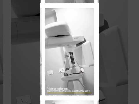

.png)


.png)

Share this listing