Restoring a missing tooth often requires more than just placing a standard implant fixture. Many patients experience significant jawbone deterioration after losing a tooth, necessitating a robust foundation through a block dental bone graft and implant procedure in Turkey. This advanced restorative technique ensures that your jaw has the structural integrity required to support a permanent titanium implant.
Choosing to undergo this complex dental restoration abroad has become an increasingly popular decision. Clinics like WestDent Clinic Turkey offer advanced surgical techniques that rival or exceed the standards found in North America and Western Europe. By understanding the anatomical requirements of a block graft and the precise steps of implant placement, patients can make highly informed decisions about their oral health investments.
Video Chapters & Quick Navigation
Understanding the Need for Bone Grafting Before Implants
When a tooth is extracted or lost to trauma, the jawbone in that specific area no longer receives the stimulation it needs from daily chewing. Without this mechanical stress, the body begins to resorb the bone tissue, using those minerals elsewhere. Within just the first year of tooth loss, a patient can lose up to twenty-five percent of their supporting bone volume.
This rapid bone resorption presents a massive challenge for oral surgeons planning a dental restoration. A standard titanium implant requires a certain width and height of dense bone to anchor properly. If an implant is placed into a deteriorated jaw, the failure rate increases drastically.
To counteract severe bone loss, surgeons utilize a block bone graft. Unlike particulate grafts, which consist of crushed bone powder used for minor defects, a block graft involves transferring a solid, square piece of bone. This provides immediate structural width and volume, making it the preferred method for major restorative cases.
Diagnosing Bone Deficiencies
Before any surgical intervention begins, dental specialists must evaluate the extent of the bone loss. This is accomplished using a 3D Cone Beam Computed Tomography (CBCT) scanner. The scan provides a highly detailed, three-dimensional view of the patient's oral anatomy.
Surgeons measure the exact millimeter dimensions of the remaining alveolar ridge. They also map out vital structures, such as the inferior alveolar nerve in the lower jaw and the sinus cavities in the upper jaw. This precise planning eliminates guesswork and ensures a safe surgical environment.
The Block Graft Harvesting Process
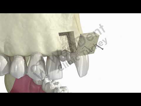
The surgical procedure begins with the careful preparation of the recipient site. As visualized at [00:04] in the clinical animation, the surgeon exposes the area of missing bone. A specialized dental handpiece is then used to prepare the site for the incoming graft.
The block of bone used for the graft can be sourced in several ways. An autogenous block graft involves harvesting a small, square section of bone from the patient's own body, usually from the back of the lower jaw (the ramus) or the chin area. Using the patient's own bone guarantees compatibility and provides live bone cells that actively stimulate healing.
Alternatively, surgeons may use an allograft block, which is sterilized donor bone sourced from a tissue bank. This eliminates the need for a second surgical site in the patient's mouth, reducing overall surgical time and post-operative discomfort. At [00:11], the precise geometric preparation of the recipient site is demonstrated, creating a perfect fit for the new bone block.
Preparing the Recipient Bed
The recipient jawbone must be prepared to accept the new block. The surgeon often decorticates the site, meaning they drill tiny holes into the surface of the existing bone. This intentionally induces a localized bleeding response.
The blood flow from the marrow spaces carries stem cells and growth factors to the surface. These biological elements are highly necessary for the new bone block to integrate successfully with the old bone. Without this vital blood supply, the graft would simply die and fail to fuse.
Securing the Graft with Fixation Screws
Once the recipient site is prepared and bleeding, the block graft is carefully positioned. It must sit flush against the host bone without any micro-movements. Even the slightest shifting during the healing phase can cause fibrous tissue to grow between the graft and the jaw, leading to graft failure.
To ensure absolute immobility, the surgeon uses tiny titanium fixation screws. At [00:19], the video illustrates the bone block being guided into the prepared cavity. Immediately following, at [00:21], two small screws are driven through the block and directly into the underlying native bone.
These screws act like lag screws in woodworking, pulling the graft tightly against the host bone bed. The tight compression maximizes cell-to-cell contact. The surgeon may then pack particulate bone material around the edges of the block to smooth out any sharp contours and ensure a natural ridge shape.
The Role of Barrier Membranes
After the block is screwed into place, the surgical site is covered with a barrier membrane. Gum tissue grows significantly faster than bone tissue. If left unprotected, the faster-growing gum tissue would invade the grafted space, preventing bone regeneration.
The barrier membrane acts as a physical shield, keeping soft tissue out while allowing the slower-growing bone cells to populate the graft. The surgeon then carefully sutures the gum tissue over the membrane, completely submerging the graft to protect it during the long healing phase.
The Healing and Osseointegration Phase
A block bone graft requires substantial time to mature before it can support a dental implant. This healing period typically ranges from four to six months. During this time, a biological process called creeping substitution occurs.
Specialized cells called osteoclasts slowly break down the grafted bone material. Simultaneously, cells known as osteoblasts lay down brand new, living native bone in its place. The graft essentially acts as a highly structured scaffold, guiding the body to rebuild the missing jaw segment.
| Graft Material | Source | Healing Time |
|---|---|---|
| Autograft Block | Patient's own jaw or chin | 4 to 5 Months |
| Allograft Block | Human tissue bank | 5 to 6 Months |
| Xenograft Block | Bovine or Porcine bone | 6 to 8 Months |
Patients are closely monitored during this healing period. Dentists advise a strict diet of soft foods initially and warn against chewing directly on the surgical site. Pressure from hard foods or poorly fitting temporary dentures can disrupt the delicate vascular network forming within the new bone.
Titanium Implant Placement Steps
Once the CBCT scan confirms the bone block has fully integrated and achieved optimal density, the patient returns for the actual implant placement. The surgeon administers local anesthesia and gently opens the gum tissue to access the newly augmented ridge.
The first step in this secondary surgery is often the removal of the titanium fixation screws that held the block graft in place. Since their job is complete, they are backed out to make room for the larger implant fixture. The surgeon then uses a series of precision drills, gradually increasing in diameter, to create the osteotomy.
At [00:30], the animation highlights the threaded titanium implant being slowly driven into the solid bone. The implant is placed with specific torque settings to achieve high primary stability. The threads of the implant bite into the dense, newly formed bone, locking it firmly into position.
The Secondary Osseointegration
Placing the implant triggers a second round of osseointegration. The titanium surface of the implant is highly biocompatible and often treated with micro-texturing to encourage cellular attachment. Bone cells migrate directly onto the surface of the titanium threads.
Over the next three to four months, the bone fuses completely with the metal. This biological lock is what allows dental implants to withstand the immense biting forces of the human jaw without loosening or causing pain.
Abutment and Crown Restoration
After the implant is solidly fused to the jawbone, the final restorative phase begins. The surgeon uncovers the top of the implant and removes the protective cover screw. An abutment is then attached directly to the implant fixture.
The abutment serves as the physical connector between the titanium root under the gums and the visible dental crown above the gums. At [00:34], we see the metallic abutment being seated onto the implant head. This component must fit perfectly to prevent bacteria from entering the internal structures of the implant.
Finally, a highly customized dental crown is fabricated to match the surrounding natural teeth. At [00:36], the white ceramic crown slides over the abutment. By [00:44], the bite is fully restored, demonstrating a seamless integration with both the upper and lower dental arches.
Choosing the Right Crown Material
Patients undergoing dental implant procedures in Turkey have access to premium restorative materials. The most common choices for final crowns are monolithic zirconia and porcelain-fused-to-metal (PFM). Zirconia has become the gold standard due to its immense fracture resistance and highly natural, translucent appearance.
The crown is either cemented onto the abutment using specialized dental cement or secured via a tiny screw that goes through the top of the crown. Screw-retained crowns are often preferred by specialists because they can be easily removed for maintenance without destroying the restoration.
Why Choose Turkey for Complex Dental Procedures
Medical tourism for dental work has seen massive growth over the last decade. Patients seeking a block dental bone graft and implant procedure in Turkey do so because of the unmatched combination of high clinical standards and affordable pricing. The cost of complex grafting in Western nations is often prohibitive for the average patient.
Turkish dental clinics operate with modern technology, utilizing state-of-the-art 3D imaging, digital impression scanners, and in-house milling machines. Clinics heavily invest in these technologies to provide rapid, precise treatment for international patients who are visiting on a tight timeline.
Expertise at WestDent Clinic Turkey
Surgeons performing these procedures in major Turkish clinics handle a massive volume of complex implant cases daily. This high frequency builds an exceptional level of surgical expertise. A specialist at a top-tier facility performs more block grafts in a single month than many general practitioners perform in a year.
Furthermore, the strict regulations enforced by the Turkish Ministry of Health ensure that clinics maintain rigorous sterilization protocols and employ fully certified specialists. Patients receive comprehensive care packages that often include VIP airport transfers, hotel accommodations, and dedicated patient coordinators.
Post-Operative Recovery Guidelines
The success of a block bone graft relies heavily on patient compliance during the post-operative recovery period. Immediately following the surgery, localized swelling, bruising, and minor bleeding are entirely normal. Patients are prescribed a strict regimen of antibiotics to prevent infection at the surgical site.
Pain management is handled through prescription analgesics and anti-inflammatory medications. Applying ice packs to the outside of the face in twenty-minute intervals during the first forty-eight hours helps significantly reduce swelling. Smoking must be strictly avoided, as the carbon monoxide and nicotine cause blood vessels to constrict, starving the new graft of essential oxygen and leading to almost certain failure.
- Maintain a Soft Diet: Consume only liquids and very soft foods like yogurt, scrambled eggs, and smoothies for the first two weeks.
- Avoid Vigorous Rinsing: Do not spit forcefully or use a straw, as the pressure can dislodge the blood clot forming over the surgical site.
- Elevate Your Head: Sleep with an extra pillow for the first few nights to reduce blood flow to the head and minimize swelling.
- Gentle Oral Hygiene: Brush surrounding teeth carefully but completely avoid the grafted area until instructed otherwise by the surgeon.
Long-Term Care for Your New Dental Implant
Once the entire process is completed and the final crown is placed, treating the implant like a natural tooth is essential. While titanium and zirconia cannot develop cavities, the gum tissue and remaining bone surrounding the implant are still highly susceptible to bacterial infection.
Peri-implantitis is a destructive inflammatory disease that affects the soft and hard tissues around dental implants. Plaque accumulation leads to inflammation, which can eventually cause bone loss and implant failure. Preventative care is the only way to ensure the lifelong longevity of the restoration.
Patients must brush twice a day and use specialized interdental brushes or water flossers to clean under the crown and around the abutment. Regular dental check-ups every six months allow specialists to take x-rays, measure bone levels, and professionally clean hard-to-reach areas, ensuring your investment remains secure for decades.
Ready to Restore Your Smile in Turkey?
If you are struggling with severe bone loss and need a reliable solution, our partnered top-tier clinics specialize in advanced block bone grafting and implant procedures. Take the first step toward a permanent, confident smile by requesting a personalized treatment plan today.
Get Free QuoteVisual Video Transcript
[00:00 - 00:04]
The animation displays a lateral view of the upper jaw showing a completely missing tooth. The gum tissue is transparent, revealing the underlying bony structure of the maxilla and indicating a loss of bone volume in the empty space.
[00:05 - 00:13]
A high-speed dental handpiece enters the frame. The drill bit carefully cuts a perfect rectangular outline into the host bone right above the area of the missing tooth, outlining the exact dimensions needed for the recipient bed.
[00:14 - 00:18]
A dental elevator instrument is inserted into the prepared groove. It carefully pries out the existing rectangular section of native bone, leaving a hollow, uniform cavity in the jawbone ready to receive the new graft material.
[00:19 - 00:29]
A solid, square block of bone graft material is brought into the surgical field. It is pressed perfectly into the hollow cavity. Two tiny titanium fixation screws are then driven through the bone block, securing it tightly and immovably into the surrounding upper jaw structure.
[00:30 - 00:34]
After a simulated healing period, a threaded titanium dental implant is shown being screwed straight upward through the gum line, passing directly through the now-integrated bone block, establishing a strong artificial root.
[00:35 - 00:46]
A customized metal abutment is seated and secured onto the top of the titanium implant. Following this, a white ceramic dental crown is lowered onto the abutment. The jaw is shown closing, demonstrating the restored bite and perfectly aligned new tooth integrating with the surrounding natural teeth.


.png)


.png)


Share this listing