Comprehensive Guide to Hyaluronic Acid Filler Complications Management in Mexico
When seeking facial rejuvenation, many patients and practitioners encounter unexpected challenges that demand immediate and precise clinical intervention. The successful management of vascular complications from hyaluronic acid fillers in Mexico requires a profound understanding of facial anatomy and instant access to emergency rescue protocols. As the global demand for dermal fillers increases exponentially, so does the statistical probability of adverse events, ranging from mild bruising to severe tissue necrosis.
Aesthetic practitioners must shift their perspective from merely reacting to adverse events to proactively implementing preventative strategies and advanced regenerative therapies. Integrating regenerative medicine in aesthetic complications—such as the use of exosomes and mesenchymal stem cells—has fundamentally changed how we restore compromised tissue. This guide provides a deep, clinical exploration of the most effective protocols to reverse filler-induced ischemia and prevent permanent disfigurement.
Video Chapters
1. The Inherent Risks of Aesthetic Injectables
Every time a needle or cannula is introduced into the dermal or subdermal layers, there is an inherent risk of compromising vascular structures. Practitioners must recognize that absolute safety in injectables is an illusion; the risk of a complication is an ever-present clinical reality. The foundation of aesthetic safety lies in profound anatomical knowledge and the ability to identify high-risk zones where vascular compromise is most likely to occur [01:35].
Vascular complications from fillers typically arise when high-G-prime cohesive gels are either injected directly into a vessel or deposited in large volumes that compress adjacent capillary networks. Unlike minor bruising, an ischemic event rapidly deprives the tissue of oxygen and vital nutrients, initiating a cascade of cellular death that can lead to permanent disfigurement. Consequently, injectors must undergo rigorous, continuous training to handle these high-stakes emergencies effectively.
2. Key Anatomical Danger Zones for Dermal Fillers
The facial artery represents the primary anatomical landmark for potential vascular events. Emerging at the anterior border of the masseter muscle, this critical vessel ascends through various facial planes, exhibiting significant multi-dimensional variability in almost every patient. It courses through the nasolabial fold as the angular artery and terminates near the medial canthus, giving rise to essential branches that supply the lips and nose.
The nasal tip and the glabella are particularly susceptible to severe ischemic events. In the nasal tip, the arterial supply transitions into a delicate, complex capillary plexus rather than distinct, large-caliber vessels. Injecting excessive volumes of hyaluronic acid—even as little as 0.1 ml—can easily overwhelm this fragile microvasculature, leading to aggressive compression syndrome. The glabella is notoriously dangerous due to the supratrochlear and supraorbital arteries, which lack robust collateral circulation, making any ischemic event in this region highly prone to rapid and severe necrosis [06:38].
Vascular Compression vs. Direct Intravascular Injection
Vascular complications manifest through two primary mechanisms: direct intravascular injection and external vascular compression. While direct cannulation of an artery results in immediate blanching and acute ischemia, external compression is often more insidious and delayed. The hydrophilic nature of hyaluronic acid means the gel will draw in water over the initial 24 to 48 hours post-procedure, continuously expanding.
This expansion increases interstitial pressure within tight anatomical compartments, gradually compressing adjacent capillaries and venules until blood flow completely ceases. This delayed compression syndrome frequently presents as a late-onset complication. Patients may leave the clinic with excellent results, only to develop a reticulated, livedo pattern or dusky discoloration hours or even days later, necessitating immediate reassessment.
3. The Holy Trinity Protocol for Vascular Complications
When a vascular complication is identified, immediate and aggressive intervention is mandatory to prevent irreversible tissue loss. The gold standard of care for resolving these aesthetic emergencies is known as the "Holy Trinity" protocol. This life-saving protocol comprises three non-negotiable pharmacological agents that every aesthetic injector must have immediately accessible in their clinical emergency kit [04:11].
- High-Dose Hyaluronidase: The primary rescue enzyme acts by hydrolyzing the cross-links of the hyaluronic acid gel. High doses—often exceeding 1,500 to 3,000 units—must be flooded into the ischemic area using a cannula or multiple needle punctures to dissolve the filler and relieve both intravascular blockages and extravascular compression instantly.
- Potent Vasodilators: Restoring blood flow to compromised tissue requires expanding the collateral vascular networks. Phosphodiesterase type 5 (PDE5) inhibitors are the drugs of choice. While Sildenafil is widely recognized, Tadalafil is frequently preferred because it achieves peak vasodilation in as little as 15 to 30 minutes.
- Antiplatelet Agents: Any vascular injury triggers the coagulation cascade. Administering an antiplatelet agent, such as Aspirin (acetylsalicylic acid) or Clopidogrel, prevents secondary thrombotic events, maintaining the patency of the microcirculation while the hyaluronidase and vasodilators take effect.
4. Recognizing Immediate vs Late Vascular Ischemia
Aesthetic practitioners must differentiate between immediate acute ischemia and delayed vascular compromise to tailor their monitoring and intervention strategies effectively. Immediate complications are typically the result of direct arterial embolization. The practitioner will notice an instant blanching (whitening) of the skin, accompanied by severe, disproportionate pain that does not correlate with the typical mild discomfort of a standard dermal injection [05:41].
Late complications, however, represent a more complex clinical challenge. These often arise between 48 to 72 hours post-procedure due to progressive swelling and localized edema. The patient may report an increasing, throbbing pain, localized swelling, and the appearance of small pustules or a mottled, net-like discoloration. This presentation is frequently misdiagnosed as an impending herpetic outbreak or a localized skin infection, delaying essential ischemic rescue protocols.
The Weekend Complication Phenomenon
A significant operational risk factor in aesthetic medicine is the scheduling timing of the procedures. A vast number of dermal filler treatments are performed on Fridays or Saturdays, aligning with patient preferences for weekend recovery. Consequently, the critical 48-to-72-hour window for delayed complications falls precisely on Sunday or Monday, when clinics are typically closed or operating with severely reduced staff.
This "weekend phenomenon" drastically increases the severity of late-onset complications. Patients may struggle to reach their practitioner, resorting to sending low-resolution photographs via messaging apps. If the practitioner is unavailable or dismisses the symptoms as normal post-procedural bruising, the critical window for early intervention rapidly closes. By Monday, the tissue may have already transitioned from ischemia to frank necrosis.
5. The Role of Regenerative Medicine in Tissue Repair
When vascular complications progress to the point of severe tissue damage, traditional dissolution protocols are no longer sufficient. Once necrosis occurs, the clinical focus must immediately shift from resolving the occlusion to aggressively stimulating the repair and regeneration of the destroyed tissue. This is where the application of regenerative medicine in aesthetics becomes completely indispensable, utilizing biological derivatives to save the face.
Mesenchymal stem cells (MSCs) and their secreted vesicles are at the forefront of this therapeutic revolution. These multipotent cells possess a profound capacity to modulate the body's immune response, reduce catastrophic tissue inflammation, and powerfully stimulate angiogenesis—the rapid formation of new blood vessels. By introducing MSCs or their derivatives into a necrotic wound bed, practitioners can halt the progression of tissue death and initiate a rapid healing cascade [14:58].
Mesenchymal Stem Cells and Exosomes for Skin Regeneration
Exosomes represent the most advanced frontier in aesthetic regenerative medicine. These nanoscale extracellular vesicles are secreted directly by stem cells and contain a concentrated, highly potent payload of messenger RNA (mRNA), microRNA, and specific regenerative signaling proteins. Unlike whole cells, exosomes do not carry the risk of immune rejection and can easily penetrate damaged cellular membranes to deliver their regenerative instructions directly into the target host cells.
When applied to ischemic or necrotic tissue, exosomes dramatically upregulate the production of vascular endothelial growth factor (VEGF), platelet-derived growth factor (PDGF), and epidermal growth factor (EGF). This biochemical cocktail forces the rapid creation of a new capillary network, restoring oxygen and nutrient delivery to the starving tissue. Clinical application of exosome serums following a severe filler complication has been shown to reduce healing times from several months down to just 10 to 21 days.
6. Understanding Collagen Modification and Aging
To fully grasp the importance of regenerative interventions, one must understand the exact dynamics of collagen production in human skin. The structural integrity and elasticity of the dermis rely primarily on the delicate balance between different collagen subtypes. In healthy, youthful skin, Type I collagen predominates, forming robust, well-organized fibrils that provide immense tensile strength and a flawlessly smooth surface texture [17:10].
As the skin ages chronologically, or when it sustains significant localized trauma from a severe ischemic event, the natural fibroblast activity alters dramatically. In individuals over the age of 60, the production of high-quality Type I collagen diminishes significantly, and the skin begins to produce a disproportionately higher ratio of Type III collagen. Type III collagen fibrils are thinner, shorter, and highly disorganized, lacking structural integrity.
Type I vs Type III Collagen Dynamics in Scarring
The primary clinical goal of applying exosomes and growth factors is to artificially manipulate this collagen dynamic during the healing phase. When a facial wound heals naturally after necrosis, the body rushes to patch the defect using rapid, disorganized Type III collagen, resulting in a severely contracted, fibrotic scar. By introducing high concentrations of targeted regenerative factors, practitioners can force the local fibroblasts to synthesize high-quality Type I collagen instead.
This biological intervention successfully prevents the dreaded retractile scarring commonly associated with severe nasal tip or labial necrosis. The application of topical collagenase can further aid this process by enzymatically degrading the disorganized Type III fibers, creating necessary spatial volume for the exosomes to stimulate the deposition of fresh, structured Type I collagen. Meticulous management of the extracellular matrix distinguishes a catastrophic aesthetic failure from a miraculous clinical recovery.
7. Advanced Treatment Protocols for Severe Necrosis
Executing a highly successful regenerative protocol for advanced necrosis requires a methodical, aggressive, and highly structured clinical approach. The treatment always begins with meticulous surgical debridement of the necrotic eschar. All dead, devitalized, and infected tissue must be carefully removed with a scalpel or curette to expose the underlying viable wound bed. This step is critically important, as biological regenerative agents cannot penetrate or function within a solid layer of dead cells [23:04].
Once the wound is thoroughly clean and bleeding slightly—indicating viable underlying microcirculation—the regenerative agents are carefully applied. Advanced techniques such as clinical microneedling (using motorized devices like Dermapen) or fractional non-ablative CO2 lasers are utilized to create thousands of micro-channels directly into the deep dermis. These channels serve as physical delivery pathways, allowing the topical serums rich in growth factors and exosomes to penetrate deeply into the tissue architecture where the target fibroblasts reside.
Utilizing Growth Factors and Exosomes for Optimal Recovery
Specific proprietary formulations, such as Activated Growth Factors (AGF) Dermal and exosome-rich serums, provide the exact biological signaling sequences required for tissue salvage. The clinical protocol often involves applying these serums directly into the micro-channels created during the treatment session, completely saturating the wound. For severe cases of necrosis, this intensive procedure is repeated rigorously every 5 to 7 days until the wound closes.
The clinical results of this intense biological stimulation are visually and functionally dramatic. Within 48 hours of the initial application, the wound edges typically exhibit active epithelialization and cellular migration. The base of the ulcer rapidly fills with healthy, beefy-red granulation tissue, indicating robust, newly formed angiogenesis. By the third or fourth treatment session, the deep tissue defect is often completely closed, leaving a flat, pink surface that successfully avoids the need for complex surgical flaps or disfiguring skin grafts.
8. Hyperbaric Oxygen Therapy in Ischemic Rescue
In conjunction with aggressive pharmacological and regenerative protocols, Hyperbaric Oxygen Therapy (HBOT) serves as a profoundly powerful adjunctive treatment for severe vascular complications. When delicate facial tissue is deprived of its blood supply due to an occlusion, localized hypoxia accelerates rapid cellular death. HBOT involves placing the affected patient in a specialized pressurized chamber where they breathe 100% pure oxygen at elevated atmospheric pressures.
This systemic process forces oxygen to dissolve directly into the blood plasma, entirely bypassing the need for red blood cell transport, which is often blocked by microthrombi or stubborn filler emboli. The hyper-oxygenation of the plasma allows life-sustaining oxygen to diffuse deeply into the ischemic tissues, significantly extending the survival window of the compromised cells. Routine clinical protocols strongly recommend daily HBOT sessions of 60 to 90 minutes until robust granulation tissue is visible and the acute threat of expanding necrosis has safely passed.
Need Expert Assistance with Aesthetic Complications?
Connect with specialized regenerative medicine clinics and board-certified practitioners who can guide you through advanced recovery protocols and safeguard your aesthetic outcomes.
Get Free QuoteVideo Transcript
00:00 CELLSTIME CLINIQUE REGENERATIVE MEDICINE
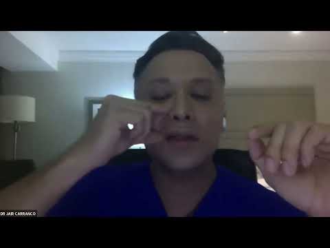
00:03 DR. JAIR CARRANCO vamos a a echar una platicadita creo que lo que vamos a ver el día de hoy que es manejo de complicaciones tardías es algo que se nos sale de pronto de las manos y que mucho tiene que ver con la inexperiencia del médico.
00:28 Me queda claro que siempre que estás inyectando tienes el riesgo de tener una complicación absolutamente siempre que estás inyectando tienes el riesgo de tener una complicación.
00:36 El detalle es que las complicaciones deberían de aprenderse a prevenir antes de trabajarlas desde esta perspectiva.
00:56 Hoy en día sabemos que tenemos complicaciones inmediatas a los procedimientos, las cuales son resultado de la práctica y de la mano que todavía no está acabada de entrenar, a veces del tipo de gel.
01:35 Sabemos que por anatomía la arteria emerge en el borde anterior del masetero y de ahí asciende y va haciendo diferentes trayectos y suele terminar en la línea de implantación capilar.
03:10 Hoy me voy a dedicar a hablar de un protocolo que nosotros hemos utilizado. Empecé a utilizar AGF como cortesía para algunos pacientes para mejorar la calidad de la piel.
04:11 La santísima trinidad de nuestra práctica es algo que tú debes de tener en tu consultorio porque no somos infalibles. Estas son: hialuronidasa, un vasodilatador (como sildenafil o tadalafil) y un antiagregante plaquetario.
05:41 Muchas veces cuando nosotros estamos inyectando con ácido hialurónico, el paciente en el momento queda bien pero entre 24 y 48 horas suele generar compresiones asociadas a la higroscopía del gel que van a disminuir el flujo.
06:38 Las complicaciones más severas regularmente tienen ciertos patrones. Hay ciertas zonas anatómicas de mayor riesgo: el ascenso de la arteria facial es el más dramático de todos. La punta de la nariz, los labios, y la glabela es sin duda la más grave.
07:18 El gran problema de estas complicaciones es que muchas se manifiestan entre viernes y sábado. Los fines de semana los pacientes buscan tratamiento y las complicaciones surgen cuando el médico no está disponible.
10:30 El colágeno en una persona joven (tipo 1) es muy diferente al colágeno que se produce después de una lesión severa o en envejecimiento (tipo 3). Necesitamos estimular la regeneración para evitar cicatrices retráctiles.
14:58 La medicina regenerativa se enfoca en estimular la regeneración de los tejidos a partir de establecer un mecanismo de inducción por factores de crecimiento, por células mesenquimales y por exosomas.
17:10 Una persona de 65 años, el colágeno que genera es principalmente tipo 3. Estas fibras son más delgadas y generan retracción de los tejidos, lo cual es vital entender cuando manejamos necrosis.
23:04 Empecé a utilizar AGF Dermal. Hacíamos curaciones, limpieza quirúrgica para hacer una debridación de todo el tejido, y al terminar poníamos un vial de colagenasa diluido para limpiar, seguido del producto regenerativo.
26:40 En solo 10 o 11 días llegamos a un cierre completo, acelerando el proceso de cicatrización y previniendo la retracción de los tejidos en la nariz. Estos casos nos dan mucha esperanza para ayudar a pacientes con complicaciones vasculares severas.


Share this listing