
Supporting Better Birth Outcomes: Intrauterine Transfusion in Saudi Arabia
Severe fetal anemia caused by maternal Anti-Kell antibodies is a complex pregnancy complication that can threaten fetal health and requires timely diagnosis, specialized monitoring, and advanced medical intervention. At King Faisal Specialist Hospital and Research Centre (KFSHRC) in Saudi Arabia, a multidisciplinary team managed a high-risk pregnancy involving a mother with a history of recurrent fetal losses over ten years. Her treatment included plasma exchange to reduce maternal antibody levels, medication, close fetal monitoring, and intrauterine blood transfusions coordinated with specialists in Riyadh. Following changes in the fetal heartbeat, an emergency cesarean delivery enabled the medical team to provide specialized neonatal care for the newborn. This case highlights the role of advanced Fetal Anemia Treatment in Saudi Arabia in managing complex fetal conditions through individualized treatment planning, coordinated specialist care, and ongoing follow-up for both mother and baby.

Quick Summary: Key Clinical Takeaways
- Critical Fetal Condition: Severe fetal anemia occurs when a fetus lacks sufficient healthy red blood cells, leading to hydrops fetalis, heart failure, or fetal loss if untreated.
- Intrauterine Transfusion (IUT): A highly specialized ultrasound-guided procedure that delivers donor red blood cells directly into the fetal umbilical vein or abdomen.
- Subspecialty Expertise: High-Risk Pregnancy Care in Saudi Arabia requires multi-disciplinary coordination across maternal-fetal medicine specialists, pediatric hematologists, and neonatologists.
- Leading Medical Center: King Faisal Specialist Hospital and Research Centre (KFSHRC) provides advanced fetal therapy and tertiary neonatal intensive care in Riyadh and Madinah.
- Early Doppler Screening: Non-invasive Middle Cerebral Artery (MCA) peak systolic velocity ultrasound is the primary diagnostic tool to assess fetal anemia severity.
- Positive Outcomes: Timely intrauterine interventions allow severely anemic fetuses to safely reach viable gestational ages for delivery.
Featured Healthcare Institution in Saudi Arabia
Real Clinical Case: Overcoming Anti-Kell Isoimmunization at KFSHRC
Patient Profile: A 34-year-old woman with a complex obstetric history of recurrent fetal losses spanning over 10 years.
Underlying Condition: Maternal Anti-Kell alloimmunization. Anti-Kell antibodies cross the placenta and destroy fetal red blood cell precursors, precipitating severe fetal anemia and hydrops.
Medical Management: Intensive therapy including maternal plasma exchange to suppress antibody titers, medication, and weekly fetal monitoring at KFSHRC Madinah.
Intrauterine Intervention: Two successful ultrasound-guided intrauterine blood transfusions coordinated closely with the specialized team at KFSHRC Saudi Arabia in Riyadh.
Delivery & Neonatal Outcome: An emergency cesarean section was performed following acute fetal heartbeat changes. A baby boy (1.65 kg) was safely delivered, receiving specialized NICU phototherapy and transfusions.
Follow-up Status: Discharged home in stable condition; multi-month follow-up confirms expected growth and normal developmental milestones with no complications.
Protecting New Life: Understanding Severe Fetal Anemia During Pregnancy
Severe fetal anemia is a life-threatening medical condition in which the circulating red blood cell count or hemoglobin concentration in an unborn baby falls significantly below normal levels. Because fetal red blood cells carry essential oxygen to developing organs, a marked reduction in oxygen-carrying capacity triggers a cascade of compensatory cardiovascular mechanisms.
Without timely diagnosis and specialized intervention, severe anemia can progress to high-output fetal heart failure, generalized fluid accumulation (hydrops fetalis), tissue hypoxia, and fetal demise. However, breakthroughs in Maternal-Fetal Medicine in Saudi Arabia now allow complex intrauterine therapies to treat fetuses directly inside the womb, protecting delicate pregnancies and leading to successful live births.
What Causes Fetal Anemia and How Can It Affect an Unborn Baby?
Fetal anemia develops through two primary physiological mechanisms: destruction (hemolysis) of fetal red blood cells or decreased fetal red blood cell production. When red blood cells are destroyed faster than the fetal bone marrow or liver can replace them, anemia ensues.
Red Blood Cell Destruction
Maternal immunoglobulin G (IgG) antibodies cross the placental barrier and target fetal red cell surface antigens (e.g., RhD, Kell, Duffy).
Suppressed Cell Production
Viral infections such as Parvovirus B19 selectively infect and destroy fetal erythroid progenitor cells, halting erythrocyte production.
Fetal Blood Loss
Placental vascular abnormalities, fetomaternal hemorrhage, or twin-to-twin transfusion syndrome (TTTS) cause direct blood loss.
Warning Signs and Risk Factors Associated With Severe Fetal Anemia
Identifying pregnant patients at elevated risk enables maternal-fetal subspecialists to initiate proactive surveillance before irreversible heart failure develops. Key risk factors and clinical warning indicators include:
- Maternal antibodies affecting fetal red blood cells: Sensitization to Kell (anti-K), RhD, RhE, or Duffy antigens from prior blood transfusions or pregnancies.
- Blood group incompatibility between mother and fetus: Rh isoimmunization occurring when an Rh-negative mother carries an Rh-positive fetus.
- Parvovirus B19 infection during pregnancy: Maternal flu-like illness or rash leading to transient fetal aplastic crisis.
- Fetal blood loss or complications involving the placenta: Placenta previa, vasa previa, or acute fetomaternal hemorrhage.
- Inherited blood disorders affecting fetal red blood cell production: Family history of severe thalassemias, enzymopathies, or red cell membrane defects.
- Conditions associated with fetal hydrops or cardiovascular strain: Ultrasound evidence of fetal scalp edema, pericardial effusion, pleural effusion, or ascites.
Global Fetal Intervention & Survival Trends in Severe Isoimmunization
Published data from leading global maternal-fetal health centers indicate a dramatic increase in overall fetal survival rates following the adoption of standardized Middle Cerebral Artery (MCA) Doppler screening and ultrasound-guided intrauterine blood transfusion protocols.
Fetal Survival Rates in Severe Alloimmunization Undergoing Intrauterine Transfusion (%)
Source: World Health Organization (WHO) & International Society of Ultrasound in Obstetrics and Gynecology (ISUOG) Fetal Therapy Guidelines Data.
Why Early Detection of Fetal Anemia Is Critical
Early detection transforms fetal anemia from a historically fatal condition into a treatable clinical scenario. Before visible anatomical abnormalities (such as abdominal ascites or cardiomegaly) appear on standard ultrasound, anemic fetuses experience hyperdynamic circulation. Recognizing these physiological changes early allows specialists to intervene prior to systemic fetal decompensation.
How Ultrasound and Doppler Studies Help Assess Fetal Anemia
The cornerstone of non-invasive fetal anemia assessment is Doppler measurement of the Middle Cerebral Artery Peak Systolic Velocity (MCA-PSV). When a fetus becomes anemic, two hemodynamic adjustments occur: blood viscosity decreases and cardiac output increases. This speeds up blood flow through the fetal brain.
The MCA-PSV Threshold Rule
An MCA-PSV value exceeding 1.5 Multiples of the Median (MoM) for a specific gestational age indicates a high probability of moderate-to-severe fetal anemia. This non-invasive screening tool has largely replaced invasive amniocentesis bilirubin testing, reducing unnecessary procedural risks.
What Is Intrauterine Blood Transfusion and When Is It Needed?
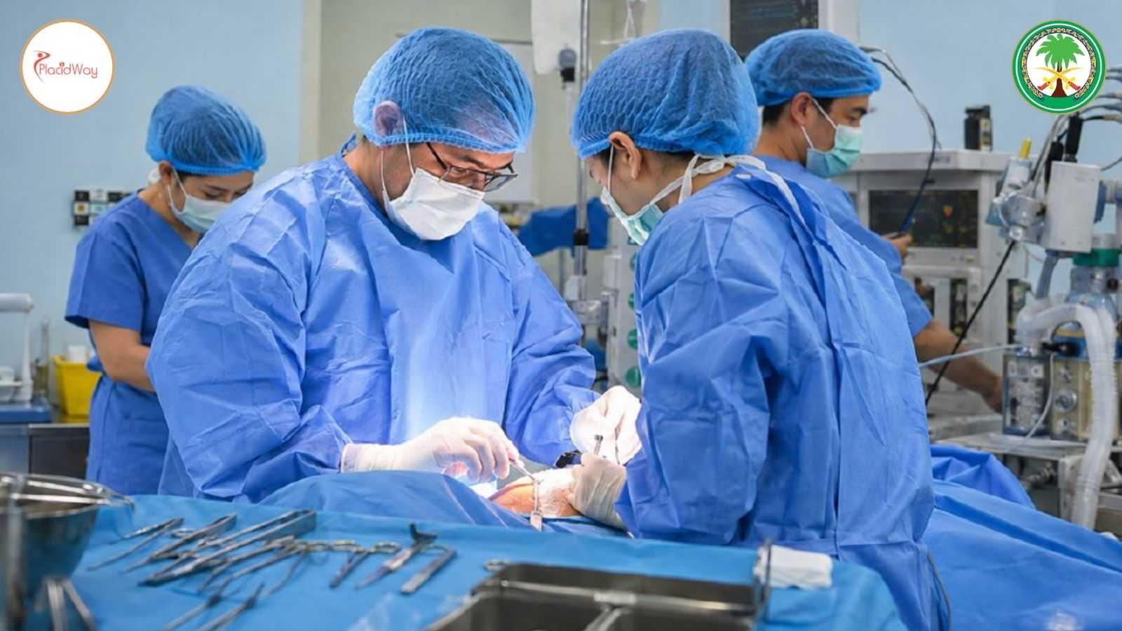
Prenatal Treatment for Fetal Anemia in Saudi Arabia relies on Intrauterine Transfusion (IUT)—a sophisticated procedure that delivers donor red blood cells directly into the fetal circulation while the fetus remains inside the uterus. It is indicated when non-invasive MCA-PSV Doppler studies or fetal blood sampling confirm severe anemia prior to 34-35 weeks of gestation.
Executing a successful IUT involves multiple meticulous clinical steps:
- Confirming the severity of fetal anemia: Serial MCA-PSV Doppler velocity calculations compared against standardized gestational curves.
- Identifying the appropriate timing for intervention: Balancing gestational age against severe anemia markers to determine optimal therapy windows.
- Determining a safe route for accessing fetal circulation: Selecting direct cord insertion access (umbilical vein at placental site) or intrahepatic vein targeting.
- Selecting compatible, specially prepared donor blood: Using O-negative, CMV-negative, irradiated, leukoreduced, tightly packed red blood cells (hematocrit 75–80%).
- Delivering red blood cells under ultrasound guidance: Real-time continuous needle monitoring by experienced maternal-fetal subspecialists.
- Monitoring fetal response during and after transfusion: Tracking intra-procedural fetal heart rate, cardiac contractility, and target post-transfusion hematocrit.
Preparing the Mother and Fetus for Intrauterine Transfusion
Pre-procedural preparation requires comprehensive maternal blood cross-matching, antibody screening, and anti-inflammatory protocols. In many instances, short-acting fetal paralytic agents are administered under ultrasound guidance to prevent fetal movement during delicate intravascular needle placement.
Key Stages of an Intrauterine Transfusion Procedure
| Procedure Stage | Clinical Actions & Objectives |
|---|---|
| 1. Pre-Procedural Review | Reviewing high-resolution fetal imaging, placental location, and maternal laboratory antibody titers. |
| 2. Needle Planning | Planning optimal ultrasound-guided needle trajectory to avoid placental disruption or cord hematoma. |
| 3. Vascular Access | Accessing the umbilical vein insertion site or intrahepatic vein with a fine 20- or 22-gauge spinal needle. |
| 4. Fetal Blood Sampling & Infusion | Drawing baseline hemoglobin, followed by slow infusion of donor packed red blood cells to reach target hematocrit (40–45%). |
| 5. Hemodynamic Monitoring | Continuous monitoring of fetal cardiac motion, heart rate, and cord insertion site following needle withdrawal. |
| 6. Post-Procedure Surveillance | Assessing need for repeat transfusions (typically required every 14–21 days until delivery clearance). |
The Role of Maternal-Fetal Medicine Specialists in High-Risk Pregnancy Care
High-Risk Pregnancy Care in Saudi Arabia relies heavily on dedicated Maternal-Fetal Medicine (MFM) subspecialists. MFM physicians undergo years of fellowship training beyond general obstetrics to master invasive in-utero surgical techniques, complex ultrasound hemodynamics, and fetal genetics.
At major medical centers like KFSHRC, MFM subspecialists direct multi-disciplinary care teams comprising fetal cardiologists, specialized blood bank hematologists, geneticists, and neonatologists to safeguard complex pregnancies.
Monitoring Fetal Well-Being After Intrauterine Transfusion
Post-transfusion surveillance ensures fetal stability and determines the precise schedule for subsequent interventions. Standard post-procedure protocols include:
- Reassessing fetal blood flow using Doppler ultrasound: Verifying that Middle Cerebral Artery blood flow velocities have normalized following red cell administration.
- Monitoring fetal heart rate and movement: Continuous cardiotocography (CTG) and biophysical profile (BPP) assessments.
- Evaluating signs of persistent anemia or fetal hydrops: Ultrasound tracking of fluid absorption in the fetal abdomen or chest cavity.
- Scheduling repeat blood tests or transfusions when indicated: Calculating donor red cell breakdown rates to time subsequent transfusions appropriately.
- Monitoring maternal health and pregnancy progression: Screening for pre-term labor, uterine contractions, or membrane ruptures.
- Planning delivery based on fetal condition and gestational age: Aiming for elective delivery between 35 and 37 weeks once pulmonary maturity is established.
Potential Benefits, Risks, and Limitations of Intrauterine Transfusion
Key Clinical Benefits
- Directly reverses severe fetal anemia and prevents in-utero heart failure.
- Resolves hydrops fetalis in up to 80–90% of cases when performed timely.
- Extends pregnancy duration to reach safe neonatal gestational viability.
- Dramatically improves overall perinatal survival rates to over 90–95%.
Procedural Risks & Considerations
- Transient fetal bradycardia or umbilical artery spasm (1–2% risk).
- Premature rupture of membranes (PPROM) or preterm labor onset.
- Umbilical cord hematoma, bleeding, or infection risks.
- Requires access to an advanced tertiary NICU in case emergency delivery occurs.
Coordinating Maternal-Fetal Treatment and Delivery Planning in Saudi Arabia
Saudi Arabia has established highly specialized tertiary referral networks connecting regional medical clinics with leading quaternary centers like King Faisal Specialist Hospital and Research Centre in Riyadh and Madinah. This integrated healthcare infrastructure allows seamless patient transfer, multidisciplinary case review, and coordinated post-delivery neonatal management.
For foreign or regional patients seeking high-risk pregnancy evaluations, establishing care at an accredited institution ensures access to state-of-the-art blood banking, continuous fetal ultrasound surveillance, and immediate Level IV Neonatal Intensive Care Unit (NICU) support upon birth.
Frequently Asked Questions
What is severe fetal anemia?
Severe fetal anemia is a critical condition where an unborn fetus has dangerously low red blood cell levels, compromising oxygen delivery to vital organs and potentially causing fetal heart failure if left untreated.
How do maternal Anti-Kell antibodies cause fetal anemia?
Anti-Kell antibodies pass through the placenta and destroy fetal red blood cells while also suppressing the bone marrow's ability to produce new red cells, leading to rapid and severe fetal anemia.
How is fetal anemia diagnosed non-invasively?
Specialists use Middle Cerebral Artery Peak Systolic Velocity (MCA-PSV) Doppler ultrasound to measure blood flow speed in the fetal brain, which accelerates when fetal blood viscosity decreases due to anemia.
What happens during an intrauterine blood transfusion?
Under continuous real-time ultrasound guidance, a physician inserts a fine needle through the mother's abdomen into the umbilical vein or intrahepatic vessel to infuse specially prepared donor red blood cells directly into fetal circulation.
How many intrauterine transfusions might a fetus need during pregnancy?
A fetus may require between 1 to 4 transfusions spaced every 2 to 3 weeks until reaching a gestational age suitable for safe delivery.
Is intrauterine transfusion safe for the mother?
Yes, intrauterine transfusion is very safe for the mother, carrying minimal maternal risks similar to standard ultrasound-guided amniocentesis procedures.
What is the survival rate for fetuses treated with intrauterine transfusion?
In specialized maternal-fetal centers like KFSHRC, overall survival rates for fetuses receiving intrauterine blood transfusion exceed 90% to 95% when intervention occurs before hydrops fetalis advances.
What treatment is needed for the newborn after delivery?
Newborns delivered after in-utero transfusions often receive specialized NICU care, phototherapy for breakdown of bilirubin, and occasionally top-up simple blood transfusions as donor cells naturally clear.
Can maternal plasma exchange reduce antibody levels during high-risk pregnancy?
Yes, maternal plasma exchange (plasmapheresis) combined with intravenous immunoglobulin (IVIG) therapy can significantly lower circulating maternal antibody titers to delay the onset of severe fetal anemia.
Why choose King Faisal Specialist Hospital and Research Centre (KFSHRC) for high-risk fetal care?
KFSHRC is a leading medical institution in Saudi Arabia featuring highly trained Maternal-Fetal Medicine specialists, advanced fetal surgery capabilities, comprehensive blood banking, and world-class Level IV neonatal intensive care units.
Connect With Maternal-Fetal Specialists at KFSHRC
If you or a family member are navigating a high-risk pregnancy involving fetal anemia, Rh isoimmunization, or complex fetal conditions, expert medical consultation is available. Request personalized details regarding Maternal-Fetal Medicine in Saudi Arabia options, medical travel coordination, and specialist evaluations.
Request Hospital InformationMedical Disclaimer: This article is published for educational and informational purposes only and does not constitute medical advice, diagnosis, or treatment. Pregnant patients experiencing high-risk conditions or suspected fetal anemia should consult immediately with a qualified Maternal-Fetal Medicine specialist or obstetrician. Treatment plans and procedural outcomes vary based on individual medical histories and clinical circumstances.
References:
- World Health Organization (WHO) — Global Maternal and Newborn Health Guidelines
- Royal College of Obstetricians and Gynaecologists (RCOG) — Management of Red Cell Antibodies in Pregnancy
- Society for Maternal-Fetal Medicine (SMFM) — High-Risk Pregnancy Interventions & Protocols
- Centers for Disease Control and Prevention (CDC) — Pregnancy Complications and Fetal Health Surveillance
- Ministry of Health Saudi Arabia — Tertiary Medical Services & Specialized Care Standards
- Fetal Medicine Foundation (FMF) — In-Utero Therapy and Fetal Anemia Doppler Assessment Standards























Share this listing