
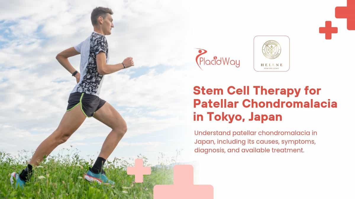
Orthopedic & Regenerative Sports Medicine Guide
Pain Relief Insights: Patellar Chondromalacia Treatment in Japan
When a sharp ache behind your kneecap turns daily stairs, desk work, and morning runs into constant frustration, understanding the root cartilage damage is the first step toward lasting relief.
The Daily Struggle Behind Knee Pain
When Every Step Downstairs Feels Like Grinding Glass
Running is celebrated as one of the most accessible ways to stay fit, clear your mind, and build cardiovascular endurance without needing expensive equipment. Yet for thousands of dedicated athletes and casual joggers alike, that freedom comes to an abrupt halt when an insistent, dull ache begins radiating from the front of the knee. What starts as mild stiffness after a weekend jog quickly escalates into sharp twinges when descending stairs, crouching to tie shoelaces, or standing up after sitting at an office desk for an hour.
For patients living with this persistent discomfort, the emotional toll can be just as heavy as the physical limitation. Watching training milestones slip away while feeling uncertain about whether resting will actually heal the joint—or whether every stride is quietly wearing away protective tissue—leaves many people stuck in a cycle of temporary ice packs, painkillers, and recurring flare-ups. While medical professionals frequently group running-related knee complaints under the umbrella term “runner’s knee,” one specific underlying condition deserves urgent attention: patellar chondromalacia.
In this comprehensive clinical guide, we break down the exact mechanics of patellar chondromalacia, how to recognize its warning signs early, why structural imbalances trigger cartilage softening, and how modern therapies—including advanced regenerative options like Stem Cell Therapy for Patellar Chondromalacia in Japan—are helping patients protect their mobility and return to active living.
Understanding the Condition
What is patellar chondromalacia?
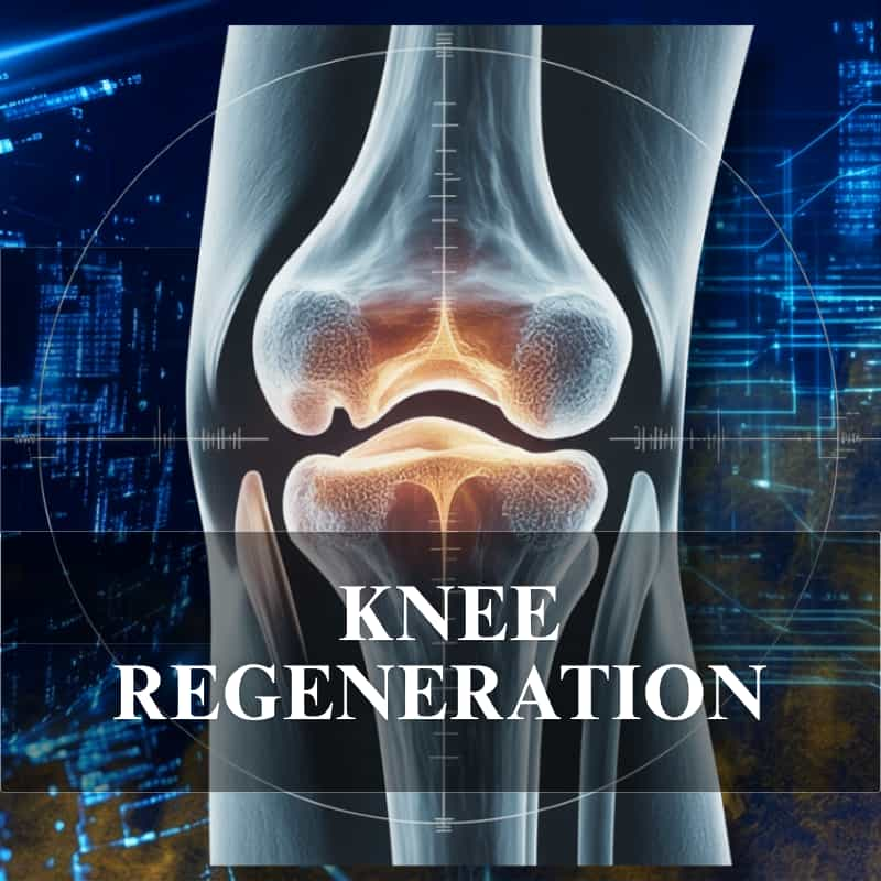
Patellar chondromalacia—often referred to clinically as chondromalacia patellae—is an orthopedic condition characterized by the gradual softening, fraying, and structural breakdown of the articular cartilage lining the underside of the kneecap (the patella). Healthy patellar cartilage acts as a smooth, resilient shock absorber that lets the kneecap glide effortlessly over the thigh bone (femur) every time you bend or straighten your leg.
Even though the affected cartilage sits on the posterior surface of the kneecap, its anatomical interface with the femoral groove means patients experience discomfort directly across the anterior (front) aspect of the knee. When repetitive friction or abnormal tracking irritates this cushion, the once-glossy cartilage loses its firmness, creating rough patches that trigger pain, stiffness, and joint inflammation.
Recognizing Early Warning Signs
Symptoms
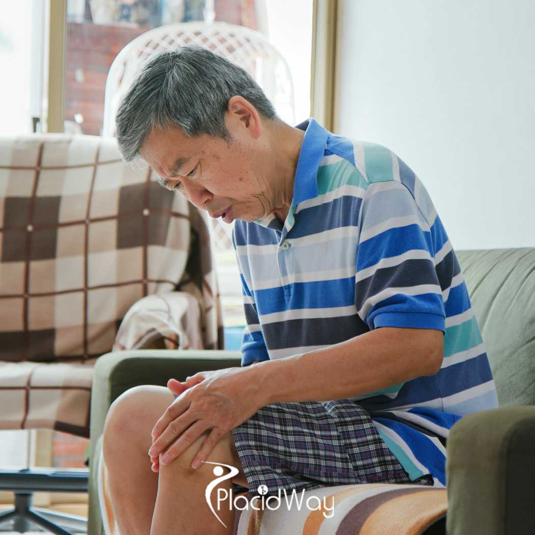
The hallmark clinical indicators of patellar chondromalacia revolve around localized anterior knee pain, subtle joint swelling, and mechanical sensations of friction during movement.
Pain
Discomfort typically concentrates directly behind, around, or immediately beneath the kneecap. During the earliest stage, soreness may surface only mid-run or during cool-down stretches. Left unaddressed, however, that ache begins intruding into routine daily movements.
Many patients notice a distinct grinding, grating, or mild catching sensation (known as crepitus) inside the joint when flexing or extending the leg—a direct result of softened, irregular cartilage surfaces rubbing against the femur.
Everyday actions that load a bent knee intensify the ache: deep squats place heavy compressive forces on the patella, and pushing upward out of a squat triggers peak contact pressure. Walking down staircases or steep hills frequently sparks sharp anterior pain, while rising from a chair after prolonged sitting (the “theater sign”) causes stiffness because inactivity temporarily reduces the circulation of protective synovial fluid across the cartilage surface.
Swelling (joint effusion)
Alongside anterior soreness, mild puffiness or fullness often develops around the perimeter of the kneecap. Unlike acute infectious inflammation marked by bright redness and intense heat, this swelling usually reflects excess synovial fluid secreted by the joint lining in response to underlying cartilage irritation—a phenomenon patients often describe as “water on the knee.”
As internal fluid volume builds, intra-articular pressure rises, producing a tight, heavy sensation that restricts full knee flexion and spreads generalized aching throughout the entire joint capsule.
Biomechanical & Training Triggers
Causes of patellar chondromalacia
Biomechanically, each footstrike during running transmits impact forces equal to roughly two to three times your total body weight directly through the knee joint. Over thousands of strides, repetitive compressive micro-trauma accumulates within the retro-patellar cartilage. Yet consistent running alone does not automatically doom your knees—rather, specific training errors, anatomical alignments, and muscular asymmetries tip the balance toward cartilage breakdown.
A sudden increase in training volume
Abrupt spikes in mileage, hill sprints, or weekly workout frequency overwhelm cartilage recovery capacity. Jumping overnight from a single 5 km jog per week to three 10 km sessions subjects the kneecap to unrelenting overload before tissue remodeling can keep pace.
Articular cartilage thrives on progressive adaptation: when training loads scale gradually over weeks, chondrocytes strengthen the extracellular matrix to tolerate higher mechanical stress safely.
Problems with physical structure
Individuals with a wider Q-angle—the structural angle formed between the quadriceps pull and the patellar tendon—experience greater lateral pulling forces that drag the kneecap slightly off-center, concentrating friction on one side of the cartilage.
Foot arch mechanics play an equally pivotal role. Collapsed arches or flat feet (overpronation) cause the lower leg to rotate inward upon landing, twisting knee alignment and creating uneven shear stress beneath the patella. Custom orthotic insoles and stability footwear often help correct these congenital alignment quirks.
Muscle balance issues
Optimal knee tracking depends on coordinated strength across the entire kinetic chain rather than isolated thigh power. When dominant quadriceps overpower underactive gluteal muscles, the knees drift excessively forward during stance phase, magnifying compressive loads against the patella.
Specific weakness in the inner quadriceps head—the vastus medialis obliquus (VMO)—impairs smooth medial patellar stabilization, while a weak core undermines pelvic control during fatigue, forcing the knees to absorb erratic torsional strain.
Differential Diagnosis for Runners
Runner’s knee in a broad sense
Clinically, “runner’s knee” is not a single diagnosis—it serves as a broad umbrella label covering several distinct overuse injuries triggered or aggravated by distance running. Alongside patellar chondromalacia, five other conditions frequently fall under this category:
| Condition | Primary Pain Location & Clinical Characteristics |
|---|---|
| Iliotibial band syndrome | Often cited as the classic form of runner’s knee; friction along the outer thigh tendon produces sharp, burning pain on the lateral (outside) aspect of the knee joint. |
| Patellofemoral pain syndrome | Presents as diffuse aching around or behind the kneecap due to tracking dysfunction, frequently overlapping with early patellar chondromalacia before visible cartilage softening occurs. |
| Patellar tendinitis | Commonly called “jumper’s knee,” this micro-tearing of the tendon connecting the kneecap to the shinbone causes focal tenderness directly below the lower pole of the patella. |
| Goose anserinus bursitis | Inflammation of the fluid-filled bursa on the upper inner shinbone produces localized pain and tenderness along the medial (inside) knee roughly two inches below the joint line. |
| Meniscus tear | Damage to the C-shaped shock-absorbing cartilage wedges inside the knee causes joint-line pain, clicking, or locking that worsens with pivoting and repetitive running impact. |
Clinical Imaging & Staging
Diagnosis of patellar chondromalacia
Accurate diagnosis combines a targeted physical examination of patellar tracking with advanced diagnostic imaging to rule out meniscal or ligamentous injuries. High-resolution Magnetic Resonance Imaging (MRI) serves as the gold-standard non-invasive tool, revealing early cartilage softening through subtle shifts in tissue thickness and internal signal intensity. High-frequency musculoskeletal ultrasound also helps clinicians evaluate cartilage contour irregularities and dynamic joint effusion in real time.
In complex or surgical cases, diagnostic arthroscopy allows direct visual inspection of the joint surface, enabling specialists to grade cartilage degeneration using the standardized Outerbridge classification system (ranging from Grade 0 for normal healthy cartilage to Grade 4 for full-thickness cartilage loss exposing underlying subchondral bone).
First-Line Conservative Care
Treatment of patellar chondromalacia
For patients diagnosed in the mild-to-moderate stages, structured conservative protocols form the cornerstone of effective Patellar Chondromalacia Treatment in Japan and worldwide, calming acute inflammation while correcting the biomechanical flaws that overloaded the kneecap.
Rest your knees
During acute pain flare-ups, pause running, deep squats, and stair-climbing workouts for 2 to 3 days to let irritated tissues settle. Strict bed rest is unnecessary—gentle walking during normal daily routines maintains healthy circulation without overloading the joint.
Once baseline pain subsides, reintroduce low-impact activity gradually over 2 to 4 weeks, stepping back immediately if sharp anterior twinges return.
Rehabilitation
Targeted physical therapy reduces patellofemoral contact stress by pairing flexibility drills with progressive muscular stabilization. Gentle quadriceps and hip-flexor stretches relieve downward tension pulling on the kneecap.
Simultaneously, isolating the vastus medialis (VMO), gluteus medius, and deep core stabilizers realigns lower-limb mechanics. Working under a qualified physical therapist ensures exercises build strength safely without aggravating existing cartilage sensitivity.
Drug therapy
When pain interferes with rehabilitation or daily mobility, clinicians may prescribe short courses of anti-inflammatory analgesics or administer intra-articular hyaluronic acid injections to restore joint lubrication, cushion shock absorption, and calm synovial irritation.
When Conservative Care Falls Short
Surgery
While the majority of individuals recover without entering an operating room, surgical intervention may be evaluated when severe structural cartilage defects fail to respond after months of dedicated Runner's Knee Treatment in Japan or specialized orthopedic rehabilitation.
Arthroscopic surgery
Keyhole arthroscopic surgery remains the primary surgical approach. Using a miniature fiber-optic camera and precision instruments inserted through tiny incisions, the surgeon trims loose, frayed cartilage flaps (chondroplasty) to create a smoother gliding surface and may perform microfracture—creating tiny perforations in the underlying bone to stimulate reparative fibrocartilage growth.
Next-Generation Biologic Cartilage Restoration
Regenerative Medicine
For active patients seeking alternatives to invasive joint surgery or long-term pain medication, regenerative orthopedics represents one of the most promising frontiers in modern Knee Joint Treatment in Japan. Rather than merely masking symptoms, biologic therapies harness the body’s intrinsic cellular repair pathways to support cartilage preservation and tissue recovery.
Autologous chondrocyte transplantation
This two-stage cellular technique involves harvesting a small biopsy of the patient’s healthy articular cartilage, expanding those chondrocytes in a specialized laboratory over several weeks, and re-implanting the multiplied cells directly into the focal patellar cartilage defect.
Stem cell therapy
Mesenchymal stem cells derived from the patient’s own adipose (fat) tissue or bone marrow possess remarkable paracrine signaling and chondrogenic potential. When processed under strict regulatory standards and delivered into the knee joint, Stem Cell Therapy in Japan aims to modulate chronic intra-articular inflammation and encourage hyaline-like cartilage matrix repair.
Platelet-rich plasma (PRP) therapy
PRP therapy concentrates autologous platelets from a simple blood draw, creating an injectable serum rich in bioactive growth factors and cytokines that help calm joint inflammation, slow ongoing cartilage degeneration, and stimulate local tissue healing.
Featured Regenerative Orthopedic Provider & Credentials
Specialized Stem Cell Care in Tokyo, Japan

Helene Clinic, Tokyo, Japan
A premier regenerative medicine facility in Tokyo specializing in autologous mesenchymal stem cell protocols for orthopedic joint preservation and sports recovery.
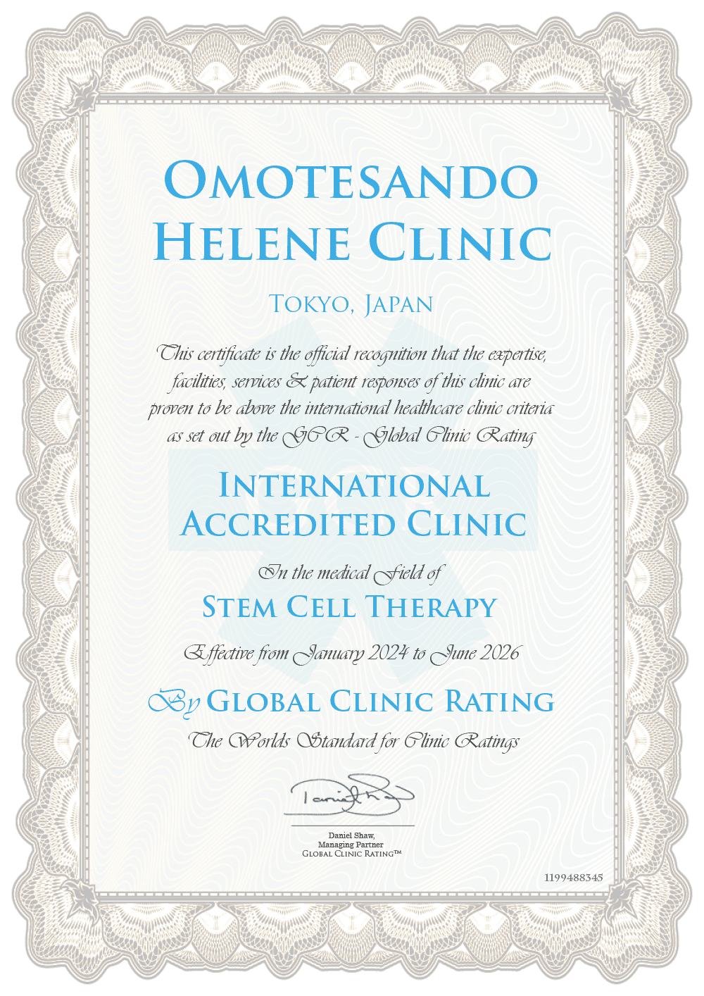
Helene Clinic Certificate
Officially licensed under Japan’s Act on the Safety of Regenerative Medicine, ensuring rigorous cell-processing quality, clinical governance, and patient safety standards.
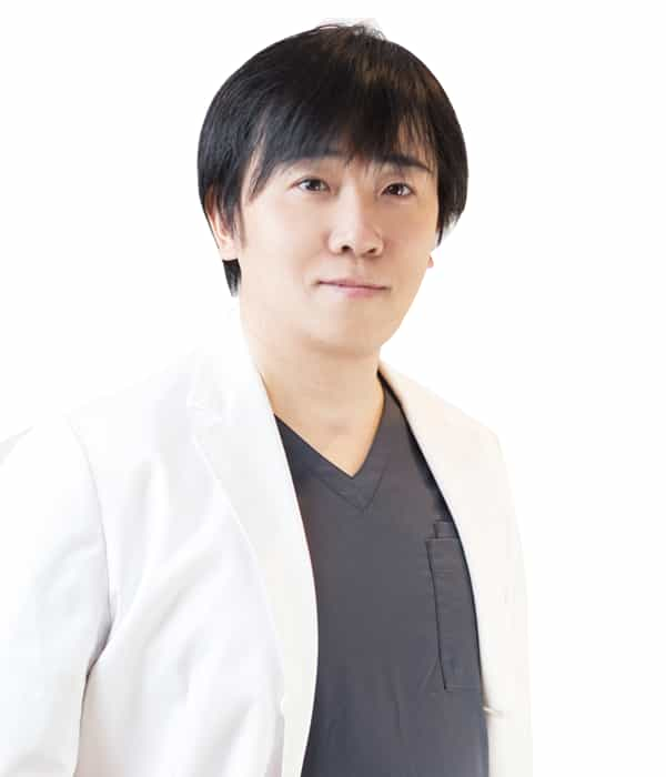
Dr. Takaaki Matsuoka - Group CEO and Chief Medical Officer
Leading clinical innovation and personalized regenerative treatment planning for international patients seeking evidence-based orthopedic and cellular therapies.
Daily Joint Protection Habits
Other measures
Long-term cartilage protection extends well beyond clinical appointments. Adjusting everyday lifestyle habits and running gear dramatically lowers cumulative stress across your kneecaps.
Weight control
Because running multiplies body weight two- to threefold at the knee, shedding even a few excess kilograms removes substantial repetitive load from patellar cartilage. Combine balanced nutrition with low-impact cross-training such as swimming or cycling.
hydration
Articular cartilage is composed largely of water held within a proteoglycan matrix, and synovial fluid relies on systemic hydration to maintain optimal viscosity. Drinking water steadily throughout the day supports smooth joint lubrication.
Choosing the Right Shoes
Properly fitted running footwear matched to your foot width and arch profile absorbs road shock before it reaches your knees. Replace worn trainers every 500–800 km—or as soon as outsole tread grooves flatten—since compressed midsoles lose shock-attenuating capacity.
Interactive Patient Q&A
Frequently Asked Questions About Patellar Chondromalacia
Click any question below to reveal clinical answers and direct guidance for managing runner’s knee and cartilage recovery.
1. Can patellar chondromalacia heal completely on its own without medical treatment?
In very mild Grade 1 stages where cartilage is only slightly softened without structural tearing, resting the joint and correcting muscle imbalances can stop further irritation and eliminate pain. However, mature articular cartilage lacks a direct blood supply, meaning deeper structural defects rarely regenerate fully without targeted clinical support.
Ask a Specialist About Your Cartilage Grade ?2. How do I tell the difference between patellar chondromalacia and patellar tendinitis?
Patellar chondromalacia causes diffuse pain behind or underneath the kneecap itself, often accompanied by grinding sensations and stiffness after sitting. Patellar tendinitis produces pinpoint tenderness directly on the tendon cord below the kneecap that hurts most during explosive jumping or sprinting.
Request an Orthopedic Diagnostic Review ?3. Why does my knee hurt more when walking down stairs than walking up?
Descending stairs requires eccentric contraction of your quadriceps while the knee bends under full body weight, compressing the softened underside of the kneecap forcefully against the femur.
Explore Joint-Preserving Treatment Options ?4. Is it safe to keep running if I only feel mild aching at the start of my jog?
Pushing through anterior knee pain—even if it temporarily warms up mid-run—accelerates mechanical friction on compromised cartilage. Taking a brief rest window and cross-training with low-impact exercise prevents mild softening from progressing into severe cartilage erosion.
Get Personalized Return-to-Running Guidance ?5. Which specific muscles should I strengthen to protect my kneecap?
Focus on the vastus medialis obliquus (inner quadriceps), gluteus medius and maximus (hip abductors and extensors), and deep core stabilizers. Balanced hip and thigh strength keeps the kneecap centered within the femoral groove during every stride.
Discuss Comprehensive Knee Rehabilitation ?6. How does stem cell therapy help patients with patellar chondromalacia?
Autologous mesenchymal stem cells release anti-inflammatory signaling molecules that calm painful joint effusion while stimulating local chondrocyte activity to support healthier cartilage matrix repair—offering a minimally invasive alternative to surgical debridement.
Inquire About Stem Cell Protocols in Tokyo ?7. What is the difference between PRP therapy and stem cell therapy for knee cartilage?
PRP delivers concentrated blood platelets rich in growth factors to reduce inflammation and relieve mild-to-moderate symptoms, whereas stem cell therapy introduces live regenerative progenitor cells capable of deeper tissue signaling and structural cartilage support.
Compare Biologic Knee Therapies ?8. Why do international patients travel to Tokyo, Japan for regenerative knee therapy?
Japan regulates regenerative medicine under strict national safety legislation (the Act on the Safety of Regenerative Medicine), ensuring certified cell-culturing facilities, high cell viability standards, and physician-supervised protocols.
Learn About International Patient Care in Tokyo ?9. What does the Outerbridge classification mean on my knee MRI report?
The Outerbridge scale grades cartilage wear from 0 to 4: Grade 1 indicates cartilage softening and swelling; Grade 2 shows partial-thickness fissures under 1.5 cm; Grade 3 reflects deep fissuring reaching subchondral bone; and Grade 4 denotes exposed bone.
Submit Your MRI Findings for Specialist Review ?10. How often should I replace my running shoes to prevent kneecap overload?
Most running shoes lose significant midsole shock absorption after 500 to 800 kilometers (roughly 300 to 500 miles) of use, or whenever outsole tread grooves appear visibly worn down.
Connect With a Knee Care Coordinator Today ?




















Share this listing