The Complete Guide to Chondromalacia: Understanding and Treating Cartilage Weakness in the Knee
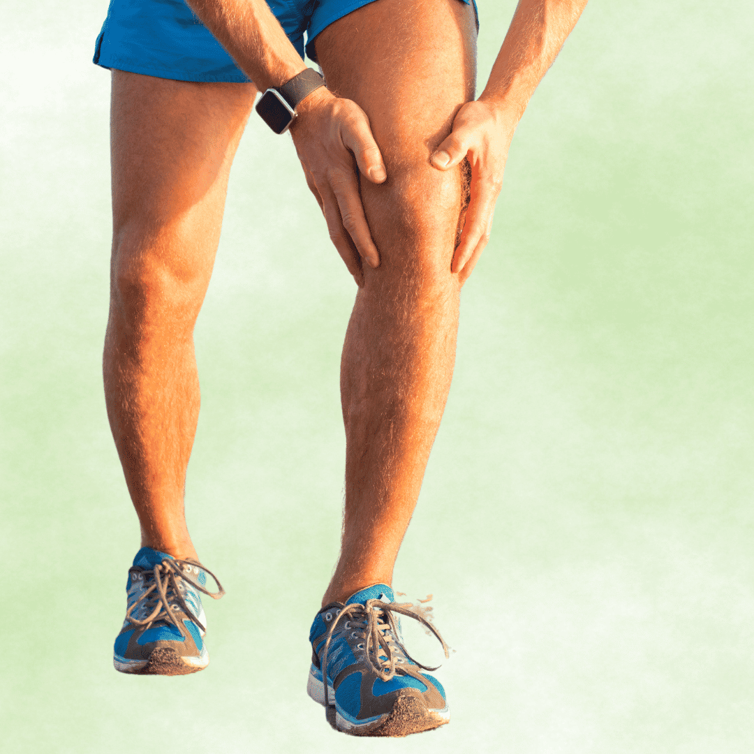
Experiencing a sharp, grinding pain behind your kneecap when climbing stairs is a warning sign you should never ignore. This discomfort is often the primary symptom of chondromalacia - cartilage weakness in knee joint, a progressive condition that breaks down the smooth tissue protecting your bones. Left untreated, this patellofemoral pain syndrome can rapidly deteriorate into permanent osteoarthritis. Understanding how to heal damaged knee cartilage is critical for restoring your mobility and preserving your joint health for years to come.
Video Chapters & Quick Navigation
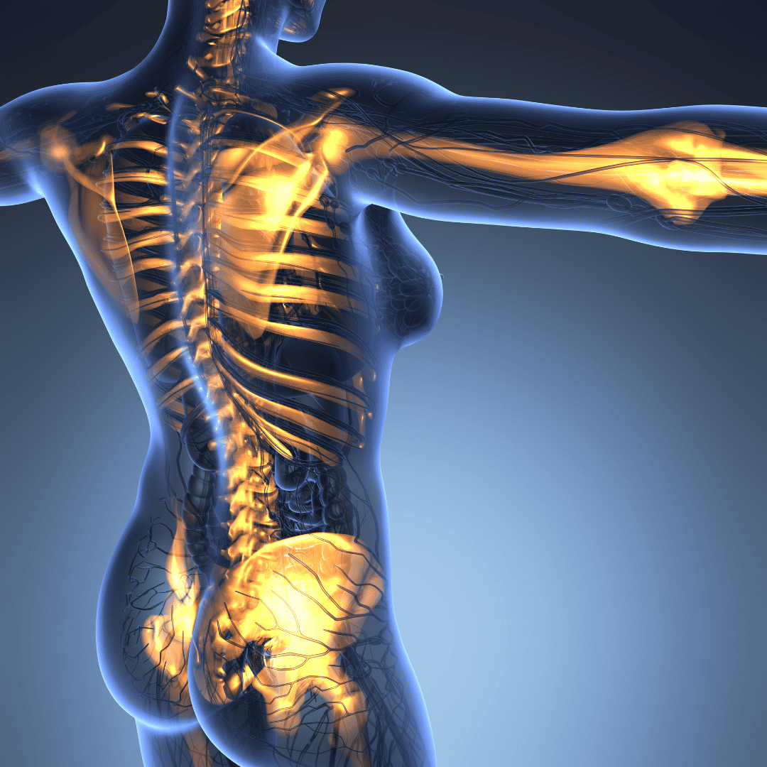
Understanding Chondromalacia Patellae and Joint Anatomy
To truly grasp the impact of chondromalacia - cartilage weakness in knee joint, one must first look at the intricate anatomy of the human knee. The knee is a complex hinge joint where the femur (thigh bone) meets the tibia (shin bone), protected at the front by the patella (kneecap). The underside of this kneecap is lined with articular cartilage, a smooth, glistening, white substance that allows the bones to glide effortlessly against one another.
When this articular cartilage begins to soften, fray, or degenerate, the condition is clinically referred to as chondromalacia patellae. This degradation means the natural shock absorption mechanism of the knee is compromised, leading directly to friction between the underlying bones. This bone-on-bone friction triggers severe inflammation within the joint capsule, creating the characteristic deep, aching pain patients experience.
Unlike muscle tissue, articular cartilage lacks a direct blood supply, which severely limits its ability to heal naturally once damaged. This biological limitation is why early intervention is absolutely vital when addressing cartilage wear and tear in the knee. Without prompt lifestyle adjustments and medical guidance, the structural breakdown will inevitably accelerate over time.
The Biomechanics of Patellofemoral Tracking
The human kneecap does not simply float in place; it is tethered securely by the quadriceps tendon above and the patellar ligament below. During normal movement, the patella is supposed to glide smoothly up and down within a specific V-shaped groove at the end of the femur, known as the trochlear groove. This precise mechanical movement is known as patellofemoral tracking.
If the muscular forces pulling on the kneecap are unbalanced, the patella can be pulled off-center, causing it to grind against the hard outer edges of the trochlear groove. This chronic maltracking applies immense, localized pressure to the fragile articular cartilage. Over thousands of steps, this uneven pressure literally grinds the cartilage away, accelerating the softening cartilage under the kneecap.
Correcting this tracking issue is the cornerstone of treating patellofemoral pain syndrome effectively. By addressing the root biomechanical flaw, patients can significantly reduce the excessive joint reaction forces that destroy cartilage during high-impact activities.
The Primary Causes of Cartilage Weakness in the Knee Joint
The breakdown of knee cartilage rarely occurs overnight; it is typically the result of cumulative stress, genetic predisposition, or acute trauma. Overuse is arguably the most common culprit, particularly among runners, cyclists, and athletes who perform repetitive, high-impact leg movements. These repetitive stress loads gradually overwhelm the cartilage's structural integrity, leading to micro-tears and softening.
Muscle imbalances, specifically weak thigh muscles, play a massive role in the development of chondromalacia. If the inner quadriceps muscle (vastus medialis oblique) is too weak compared to the outer thigh muscles, the kneecap is naturally pulled laterally out of its groove. Poor foot biomechanics, such as flat feet or overpronation, can also alter the alignment of the entire leg, forcing the knee to rotate abnormally inwards during walking.
Direct trauma to the kneecap, such as a severe fall or a dashboard impact during an auto accident, can cause sudden and severe cartilage lesions. Additionally, natural aging causes a decrease in the water content and elasticity of cartilage, making older adults highly susceptible to joint wear and tear even from low-intensity daily activities.
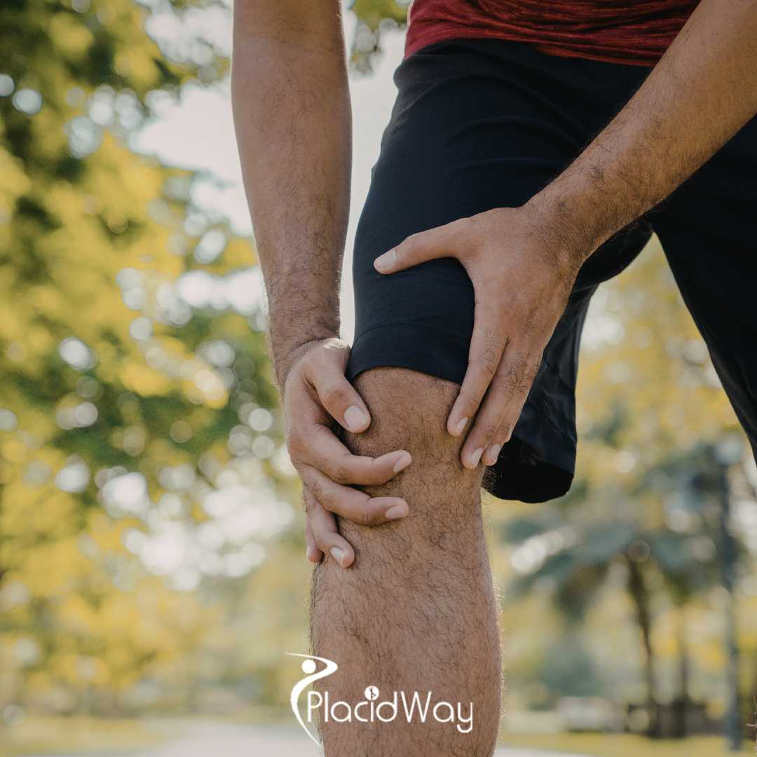
Identifying Symptoms: Knee Pain When Bending or Climbing Stairs
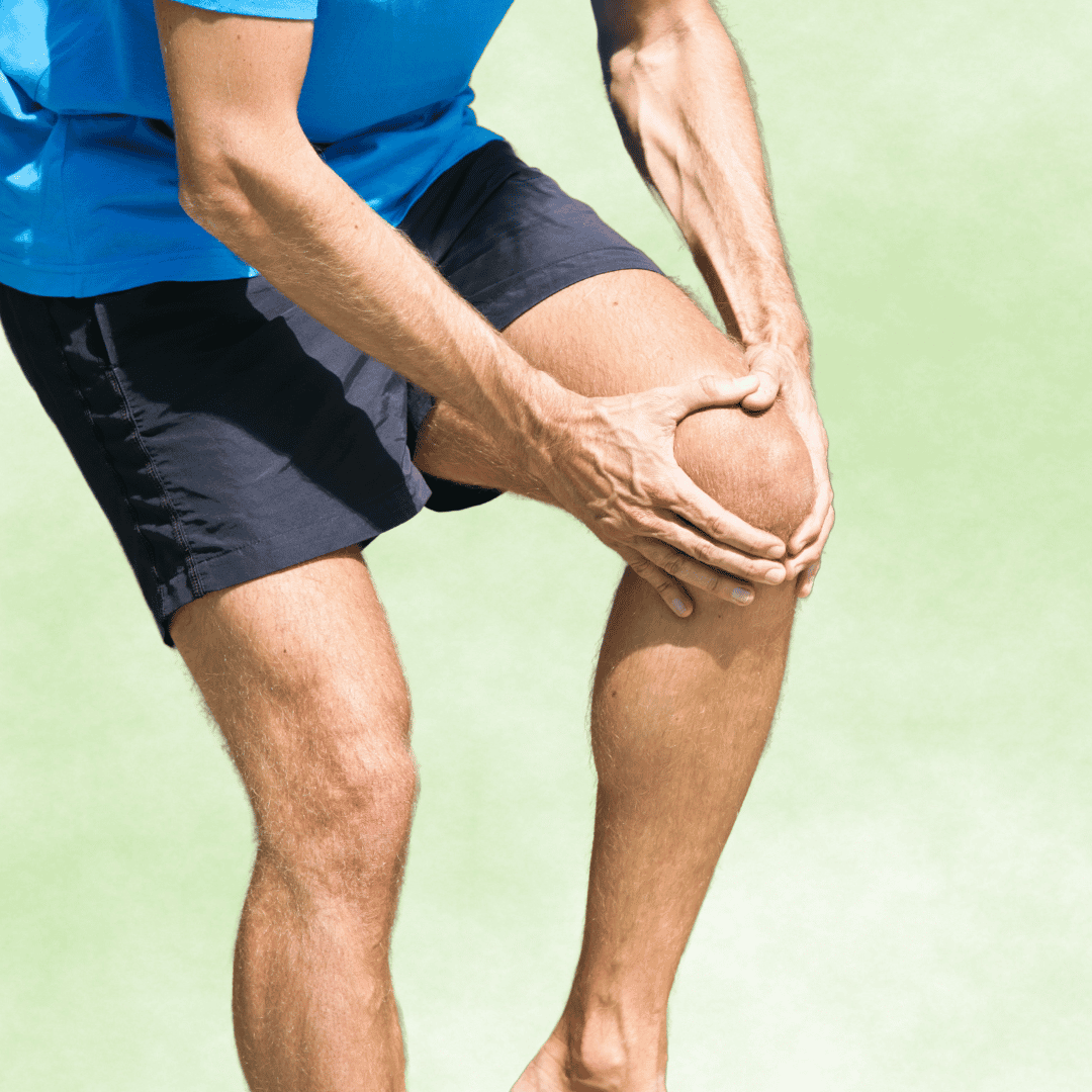
Recognizing the clinical signs of chondromalacia early can mean the difference between non-invasive physical therapy and the need for complex orthopedic surgery. The hallmark symptom is a dull, aching pain localized at the front of the knee, positioned directly behind or around the kneecap. This discomfort is heavily exacerbated by activities that load the patellofemoral joint under deep flexion.
Patients almost universally report an increase in knee pain when bending or climbing stairs. The sheer physics of ascending or descending stairs can multiply the force across the knee joint to up to four times an individual's total body weight. Squatting, kneeling, or sitting for prolonged periods with bent knees—often referred to as "theater sign" or "movie-goer's knee"—will also trigger deep joint aching.
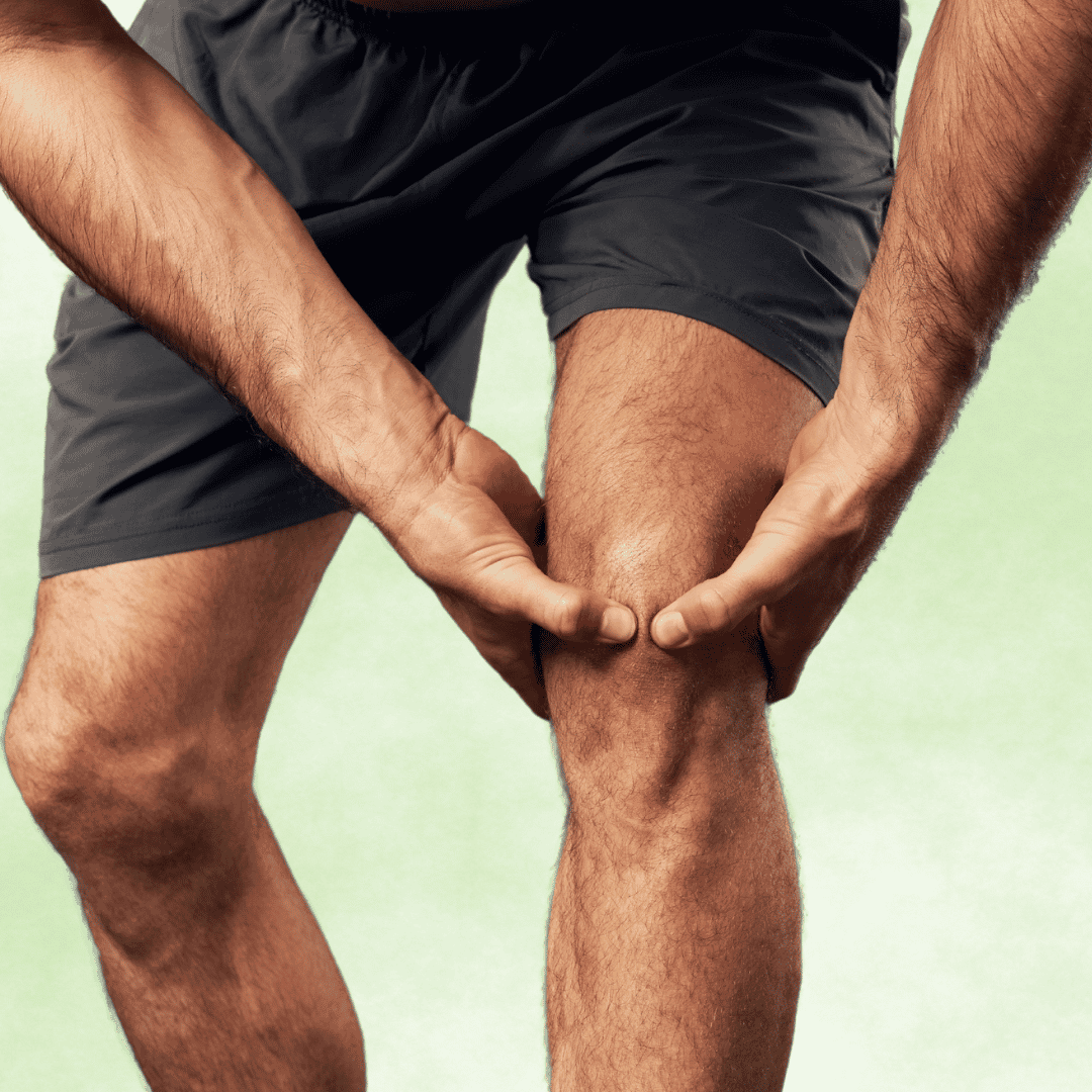
Another prevalent physical symptom is crepitus, a distinct grinding, clicking, or crunching sensation felt and sometimes heard when extending the leg. In more advanced cases, inflammation of the joint lining can lead to mild but persistent swelling, making the knee feel stiff and reducing the overall range of motion.
The Four Stages of Cartilage Wear and Tear in the Knee
Medical professionals grade the severity of chondromalacia patellae on a scale from one to four. Understanding these specific stages helps orthopedic specialists formulate an effective protocol for how to heal damaged knee cartilage.
| Stage | Medical Description | Clinical Impact |
|---|---|---|
| Grade 1 | Softening and blistering of the articular cartilage. | Mild discomfort after strenuous activities; highly responsive to rest. |
| Grade 2 | Fraying of the cartilage surface with minor tissue erosion. | Noticeable grinding sensation; pain during stair climbing. |
| Grade 3 | Deep fissures and thinning of the cartilage layers. | Significant chronic pain, joint stiffness, and frequent swelling. |
| Grade 4 | Complete cartilage loss, exposing the underlying subchondral bone. | Severe bone-on-bone friction; major loss of joint mobility. |
How Specialists Diagnose Patellofemoral Pain Syndrome
Securing an accurate diagnosis is the critical first step before engaging in any knee cartilage repair protocols. An orthopedic physician will begin with a comprehensive physical examination, carefully manipulating the knee to pinpoint the exact source of pain. They will assess the tracking of the kneecap while the patient extends their leg, looking for lateral deviations.
One common diagnostic maneuver is Clarke's test, where the doctor applies gentle downward pressure on the patella while asking the patient to contract their quadriceps. If this causes significant pain, it is a strong clinical indicator of patellofemoral cartilage irritation. The doctor will also check for tightness in the IT band, hamstrings, and calves, as inflexibility directly contributes to joint compression.
To confirm the diagnosis and rule out other issues like meniscus tears, imaging studies are usually ordered. While standard X-rays are excellent for identifying bone spurs or arthritis, they cannot show soft tissue. An MRI (Magnetic Resonance Imaging) is the gold standard for visualizing the softening cartilage under the kneecap and determining the exact grade of chondromalacia.
Conservative Treatments to Heal Damaged Knee Cartilage
For individuals presenting with Grade 1 or Grade 2 chondromalacia, non-surgical conservative treatments yield excellent success rates. The immediate goal of these interventions is to drastically reduce joint inflammation and eliminate the biomechanical stress causing the cartilage wear. This process begins with the classic RICE method—Rest, Ice, Compression, and Elevation.
Patients must temporarily halt all high-impact activities, swapping running and heavy lifting for swimming or stationary cycling. Nonsteroidal anti-inflammatory drugs (NSAIDs) may be prescribed to control the swelling within the knee capsule, providing a critical window for physical therapy to begin. In some cases, a specialized knee brace or patellar tracking tape is applied to artificially hold the kneecap in proper alignment during daily movements.
Custom orthotic shoe inserts are often highly recommended for patients whose patellofemoral pain syndrome is driven by flat feet or overpronation. By correcting the arch of the foot, orthotics successfully realign the tibia and femur, which in turn normalizes the tracking of the kneecap. Do not ignore the pain, as early conservative management is the only way to prevent severe degradation.
Essential Exercises for Chondromalacia Patellae
Physical therapy is undeniably the most crucial component of recovering from cartilage weakness in the knee joint. A targeted exercise program aims to rebuild the muscular support system around the knee without placing excessive compressive loads on the patella itself. Building strength correctly ensures the joint remains stable during dynamic movements.
Targeted Muscular Strengthening Protocols
- Straight Leg Raises: By lying flat and lifting a locked, straight leg, patients can activate the quadriceps heavily without bending the knee. This prevents the kneecap from grinding against the femur while still building essential muscle tone.
- VMO Activation: The vastus medialis oblique, the teardrop-shaped muscle on the inner thigh, is notoriously difficult to isolate. Focusing on strengthening this specific muscle corrects the lateral pull that causes maltracking.
- Clamshells and Hip Abduction: The hips act as the steering wheel for the knees. Strengthening the gluteus medius prevents the knee from collapsing inward during a walking stride, drastically reducing patellar stress.
- Hamstring Stretching: Tight hamstrings force the quadriceps to work significantly harder during a normal gait cycle. Stretching the back of the leg reduces the resting tension placed directly on the kneecap.
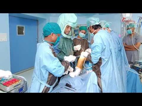
Advanced Cartilage Regeneration Knee Surgery
When conservative treatments fail to provide relief, or when a patient presents with Grade 4 bone-on-bone chondromalacia, advanced surgical intervention becomes necessary. The field of orthopedics has made massive technological leaps in cartilage regeneration knee surgery over the past decade. The primary goal of these surgeries is to clean out damaged tissue and stimulate the body’s natural healing response.
Arthroscopic chondroplasty is the most common initial surgical approach. Using a tiny camera and specialized instruments, a surgeon enters the knee joint to carefully shave away the frayed, damaged cartilage flaps. By smoothing the underside of the kneecap, the immediate mechanical friction and grinding are eliminated, offering rapid pain relief for the patient.
For highly localized areas of total cartilage loss, surgeons may perform a microfracture procedure. During a microfracture, the surgeon drills tiny holes into the exposed subchondral bone beneath the kneecap. This intentional trauma releases bone marrow and stem cells into the joint space, which then clot and form a functional, protective layer of fibrocartilage over the bare bone.
Nutritional and Medical Support for Joint Health
While physical therapy and biomechanical corrections form the foundation of recovery, optimizing the internal environment of the knee is equally important. Nutrition plays a surprisingly large role in managing chronic joint inflammation. Patients are strongly encouraged to adopt an anti-inflammatory diet rich in omega-3 fatty acids, antioxidants, and lean proteins to support overall tissue health.
Clinical studies have shown that high-quality dietary supplements can offer symptomatic relief for patellofemoral pain syndrome. Glucosamine and chondroitin sulfate are naturally occurring compounds found in human cartilage, and supplementing them may help slow the degradation process. Additionally, collagen peptides provide the essential amino acid building blocks the body needs to maintain connective tissue elasticity.
From a medical standpoint, intra-articular injections offer an excellent bridge between physical therapy and surgery. Corticosteroid injections can provide rapid, massive reductions in severe swelling. Alternatively, Hyaluronic Acid (HA) injections act as artificial joint fluid, heavily lubricating the knee joint and reducing the mechanical friction associated with cartilage wear and tear in the knee.
Long-Term Strategies for Joint Preservation
Effectively managing chondromalacia - cartilage weakness in knee joint is not a short-term fix; it requires a lifelong commitment to joint preservation. One of the most impactful long-term strategies is maintaining a healthy body mass index (BMI). Because every pound of body weight equates to up to four pounds of force on the knee during stair climbing, even a five-pound weight loss removes twenty pounds of stress from the patellofemoral joint with every single step.
Activity modification is another vital component of long-term joint health. Individuals prone to cartilage damage must learn to listen to their bodies and avoid pushing through sharp joint pain. Transitioning from high-impact sports like distance running to lower-impact activities like elliptical training or swimming allows for excellent cardiovascular fitness without sacrificing knee health.
Finally, prioritizing proper footwear cannot be overstated. Shoes that provide excellent arch support and impact cushioning absorb the shock waves that would otherwise travel directly up the shin bone and into the kneecap. By combining intelligent biomechanics, consistent physical therapy, and proactive weight management, patients can successfully halt cartilage degradation and live a highly active, pain-free life.
Ready to Treat Your Chronic Knee Pain?
Don't let worn-out cartilage dictate your lifestyle. PlacidWay Medical Tourism connects you with top-rated orthopedic specialists globally, offering world-class cartilage repair and regenerative therapies at highly affordable prices. Let us help you take the first step toward a pain-free life.
GET A FREE ORTHOPEDIC QUOTE


.png)
.jpg)


Share this listing