Living with blurred vision can severely impact your quality of life, but treating it shouldn't drain your life savings. For years, patients facing astronomical out-of-pocket costs for premium eye care have felt trapped by limited local options.
Today, affordable cataract surgery in Mexico offers a brilliant alternative. You no longer have to compromise on surgical quality to stay within budget. Watch this comprehensive surgical walkthrough to discover how the specialists at Tijuana Eye Center utilize world-class phacoemulsification technology to flawlessly restore vision, combining top-tier medical expertise with extraordinary savings.
Video Chapters & Quick Navigation
Understanding the Global Burden of Vision Loss and Cataracts
Cataracts remain the leading cause of reversible blindness worldwide, affecting millions of adults every single year. As the natural crystalline lens inside the eye begins to age, proteins cluster together, forming dense, opaque clouds that physically block incoming light. The condition is often slow and insidious, creeping up on patients over years or even decades before necessitating medical intervention.
Initially, you might notice that colors appear washed out or that driving at night becomes increasingly difficult due to excessive glare from oncoming headlights. Over time, the world simply loses its sharpness, forcing many to abandon hobbies like reading, sewing, or playing golf. When left untreated, this progressive clouding will inevitably lead to severe visual impairment.
The Psychological Impact of Vision Loss
The psychological toll of losing your visual acuity is profound and heavily documented in medical literature. Independence is closely tied to our ability to navigate the world safely without relying on the constant assistance of others. When a clouded lens prevents you from driving to the grocery store or recognizing the faces of your loved ones from across the room, the world becomes a much smaller, more isolating place.
Finding an affordable vision correction abroad is not just a financial decision; it is a critical step toward reclaiming personal freedom and mental well-being. Medical interventions are absolutely necessary to halt this decline, but the exorbitant fees charged by regional healthcare systems often act as an insurmountable barrier to entry for everyday people.
The Rise of Medical Tourism for Ophthalmic Care
Faced with deductibles that can reach thousands of dollars and insurance policies that refuse to cover anything beyond a basic, rudimentary monofocal lens, patients are taking matters into their own hands. The phenomenon of traveling for healthcare has evolved rapidly over the last decade. People are no longer seeking out questionable back-alley clinics; they are booking appointments at internationally accredited hospitals that frequently surpass the technological standards found in their own hometowns.
Securing an affordable cataract surgery in Mexico has transitioned from a niche concept into a mainstream strategy for proactive healthcare consumers. By stepping just outside their domestic healthcare borders, patients are discovering a world where premium eye care is treated as a fundamental right rather than a luxury privilege.
The Tijuana Medical Excellence Phenomenon
Tijuana, located just minutes across the border from San Diego, California, has proudly positioned itself as the undisputed capital of North American medical tourism. The geographical convenience is undeniable, but it is the stringent medical excellence that truly drives the massive influx of international patients. The top eye specialists operating at the best eye clinic in Tijuana undergo rigorous, globally recognized medical training.
It is standard practice for these elite ophthalmic surgeons to complete advanced fellowships and board certifications in the United States, Canada, or Europe. Operating rooms are equipped with the latest generation of diagnostic imaging, laser suites, and advanced surgical microscopes. By capitalizing on lower regional operational costs, these clinics can pass immense savings directly to the patient, making premium intraocular lens implantation an accessible reality.
Advanced Diagnostics: Mapping the Eye for Surgical Success
The success of premium intraocular lens implantation does not solely rely on the surgeon's steady hands; it is equally dependent on the exhaustive diagnostic testing performed long before the patient enters the operating theater. Modern ophthalmology relies heavily on digital biometry, advanced topography, and high-definition imaging. These tools allow the surgical team to create a highly customized treatment plan tailored perfectly to your unique ocular anatomy.
Precision Biometry and Ultrasound
To calculate the exact optical power required for the new IOL, technicians utilize advanced optical biometers. These non-invasive lasers measure the precise axial length of the eye, from the peak of the cornea to the macula on the retina, down to the absolute micrometer. Traditional ultrasound devices may also be utilized for patients with extremely dense cataracts that standard laser light cannot easily penetrate.
The collected ocular data is then fed into complex mathematical formulas, such as the Barrett Universal II or the SRK/T algorithms. These formulas calculate the perfect lens power necessary to hit the patient's specific refractive target, ensuring a flawless visual outcome post-surgery.
Topography and Cell Counts
Corneal topography maps the highly detailed surface curvature of the eye, generating a three-dimensional model of its microscopic hills and valleys. This is absolutely critical when determining if a patient is a suitable candidate for an astigmatism-correcting toric lens. By identifying the exact axis of the corneal irregularity, the surgeon knows precisely how to orient the implant during the procedure.
Additionally, an endothelial cell count is meticulously performed via specular microscopy. This diagnostic test ensures that the patient has a healthy reserve of inner corneal cells, guaranteeing that the eye will recover swiftly from the high-frequency ultrasonic energy used during the phacoemulsification phase.
Advanced Phacoemulsification: The Gold Standard in Lens Replacement
To truly appreciate the immense value offered by medical tourism, one must understand the absolute sophistication of the surgical technique being utilized. Phacoemulsification is a modern marvel of biomedical engineering and refined surgical skill. Gone are the days when a cataract required a massive, traumatic incision halfway across the eye, followed by weeks of painful healing and heavy visual distortion.
Today, the phacoemulsification procedure is elegantly minimally invasive, designed specifically for rapid visual recovery and minimal tissue disruption. It is this specific technological leap that makes traveling for out of pocket cost for cataract surgery such a safe, reliable, and appealing proposition for international patients.
Precision from the First Micro-Incision
The entire transformative procedure begins with the creation of a micro-incision. As clearly seen in the surgical footage at [00:02], the surgeon uses a specialized micro-keratome blade to make a tiny, self-sealing tunnel right through the clear cornea. This precise entry point is usually no larger than 2.2 to 2.4 millimeters wide, keeping the overall structural integrity of the eye intact.
Because of its unique, step-like architectural design, the natural internal pressure of the eye will effortlessly hold this incision closed once the surgery is complete. This architectural marvel vastly reduces the risk of postoperative infection and generally eliminates the need for heavy, uncomfortable sutures.
Protecting the Delicate Inner Environment
Once the access point is established, the surgeon must immediately protect the fragile internal environment of the anterior chamber. At [00:23], a thick, protective viscoelastic gel is meticulously injected into the eye. This medical-grade, honey-like substance acts as an essential shock absorber for the surrounding tissues.
It maintains the necessary volume and structural shape of the eye while providing a safe, lubricated workspace for the sharp surgical instruments. Most importantly, it shields the corneal endothelium—the critical single layer of non-regenerating cells responsible for keeping the outer cornea perfectly clear throughout your life.
Harnessing Ultrasonic Energy for Safe Extraction
At the absolute heart of the procedure is the phacoemulsification probe, which you can see deployed at [01:15]. This sophisticated titanium instrument oscillates at extreme ultrasonic frequencies, often upwards of 40,000 times per second. When brought into careful contact with the dense, clouded cataract, these microscopic vibrations effectively pulverize the hardened protein clusters.
The advanced surgical microscope and operating room setup, clearly visible at [00:56], allow the surgeon to monitor every microscopic movement with brilliant, high-definition clarity. As the ultrasound energy melts the tissue, a built-in vacuum system immediately aspirates the debris. At [02:00], you can observe the careful and methodical removal of these emulsified lens fragments, ensuring that the delicate capsular bag remains completely pristine and intact.
Following the main extraction, a specialized irrigation and aspiration (I&A) tool is smoothly introduced at [03:00]. The cortex, a softer, sticky layer of the cataract, often clings stubbornly to the inner walls of the capsule. The I&A handpiece gently vacuums away these final remnants, leaving behind a perfectly polished anatomical pocket ready to receive the artificial lens.
Navigating the Complex World of Intraocular Lenses (IOLs)
Removing the cloudy, dysfunctional lens is only the first half of the surgical equation; successfully replacing its refractive power is the critical second half. In the past, patients who underwent cataract extraction were forced to wear incredibly thick, heavy "coke-bottle" glasses just to see their surroundings. The modern invention of the Intraocular Lens (IOL) completely revolutionized global ophthalmology.
When exploring medical tourism for eye surgery, patients gain unprecedented access to the very best premium lenses that might otherwise be cost-prohibitive in their home country. To help patients understand their incredible options, here is a detailed breakdown of the premium IOLs frequently utilized by top surgical centers:
| Lens Classification | Primary Optical Function | Ideal Patient Profile |
|---|---|---|
| Standard Monofocal | Provides crystal clear vision at a single, fixed distance (usually optimized for far away). | Patients perfectly comfortable wearing reading glasses for close-up tasks like texting, cooking, or reading. |
| Premium Toric | Features customized, built-in astigmatism correction to neutralize an irregularly shaped cornea. | Individuals with pre-existing astigmatism seeking to permanently eliminate their dependence on distance glasses. |
| Advanced Multifocal | Uses concentric optical rings to split light, providing simultaneous near, intermediate, and distance vision. | Patients demanding maximum independence from all eyewear, allowing them to read, work on a computer, and drive seamlessly. |
| EDOF (Extended Depth of Focus) | Creates an elongated focal point to provide excellent intermediate to distance vision with fewer nighttime halos. | Highly active individuals who prioritize flawless intermediate vision for sports, computer work, and nighttime driving. |
The Brilliant Engineering Behind Foldable Lenses
Modern IOLs are constructed from highly advanced, biocompatible materials, primarily hydrophobic or hydrophilic acrylics. These space-age materials are incredibly flexible, which is a vital, non-negotiable characteristic for modern micro-incision surgery. The lens must be pliable enough to fold without breaking.
At [04:18], we see the surgical assistant carefully loading the selected IOL into a specialized delivery cartridge. The delicate lens is folded completely in half, much like a taco, allowing it to easily pass through an incision that is significantly narrower than the lens itself.
Step-by-Step Breakdown of the Final Surgical Phases
The meticulous preparation and exhaustive cleaning of the eye lead to the most transformative moment of the entire procedure: the physical implantation of the new artificial lens. This critical step requires absolute precision, steady hands, and a deep, intuitive understanding of fluid dynamics within the eye.
Implanting the Future of Your Vision
With the capsular bag cleaned and stabilized by viscoelastic gel, the surgeon carefully introduces the injector tip directly into the corneal micro-incision. As flawlessly demonstrated at [04:40], the tightly folded acrylic lens is slowly and smoothly injected into the eye. It is a visually mesmerizing process to witness; the lens gently unfolds within the liquid environment, gracefully spreading its haptics to anchor itself securely within the capsular equator.
Once deployed, the surgeon uses a remarkably fine instrument to dial the lens into its exact optimal position. If an astigmatism correcting toric lens is being used, it must be manually rotated to align precisely with the specific axis of the patient's corneal irregularity. Even a slight misalignment of just a few degrees can drastically reduce the refractive effectiveness of the implant.
Securing the Eye and Preparing for Recovery
Once the intraocular lens is perfectly centered, the remaining viscous material must be completely flushed out to prevent post-operative pressure spikes. The surgeon meticulously irrigates the entire anterior chamber with sterile saline. Because the original primary incision was created with a specific step-like architecture, it will naturally seal itself as the eye reaches its normal internal pressure parameters.
However, in certain specific cases, a careful surgeon may decide to place a single, microscopic stitch to ensure absolute stability, especially if the patient plans to travel shortly after. We can clearly observe this incredibly delicate suturing technique at [05:30]. This fine nylon thread is significantly thinner than a human hair and provides an extra layer of structural integrity during the crucial first week of healing.
Finally, at [06:15], the complex procedure is finalized. The eye is gently cleaned, a series of powerful antibiotic drops are applied, and a protective plastic shield is taped over the eye. The entire process, from the first precise incision to the final stabilizing suture, takes mere minutes to execute, yet its profound impact easily lasts a lifetime.
The Post-Operative Journey: Healing and Vision Restoration
One of the most universally praised aspects of modern phacoemulsification is the incredible speed at which patients recover their visual function. For many individuals, the dramatic improvement is noticed almost immediately in the recovery room. The dense, yellow-brown biological filter that had been aggressively dulling their vision for years is entirely gone, replaced by a pristine, optically perfect piece of medical-grade acrylic.
Patients routinely report that colors suddenly appear incredibly vibrant and true to life. Whites are suddenly a pure, brilliant white, and blues are deeply saturated. The profound clarity is often an emotional experience for those who had slowly forgotten what high-definition vision truly looked like.
Navigating the First 24 Hours of Recovery
The first day following the surgery is crucial for ensuring proper, uncomplicated healing. Patients are typically advised to rest comfortably and strictly avoid any strenuous physical activity, heavy lifting, or bending over at the waist, as these actions can temporarily spike the intraocular pressure. A strict daily regimen of prescription anti-inflammatory and antibiotic eye drops is provided to prevent infection and control the natural inflammatory response.
While experiencing some mild scratchiness, a slight foreign body sensation, or minor light sensitivity is entirely normal as the cornea heals, severe, throbbing pain is exceedingly rare. Patients flying back home are given explicit post-operative guidelines to ensure their travel is safe and entirely comfortable.
Long-Term Maintenance and Lifelong Eye Health
Unlike natural biological lenses, artificial intraocular lenses do not degrade, age, or cloud over time. They will absolutely never develop another cataract. The biocompatible acrylic is brilliantly designed to remain entirely clear and functional for the entirety of the patient's natural life. By simply adhering to routine, annual follow-up eye exams and maintaining a healthy lifestyle, patients can comfortably enjoy their newfound visual clarity indefinitely without the fear of recurring cataracts.
The Cost Analysis: Tijuana vs. The United States and Canada
The fundamental driving force behind the global medical tourism boom is the undeniable, deeply frustrating discrepancy in healthcare economics. The out of pocket cost for cataract surgery in the United States has violently skyrocketed over recent years. Even with the assistance of Medicare or private health insurance, patients are routinely hit with substantial, unexpected copays, massive facility fees, and separate anesthesiologist bills.
More importantly, domestic insurance plans rigidly classify premium IOLs—such as highly desirable multifocal or toric lenses—as completely "elective" or "cosmetic" luxury upgrades. This bureaucratic classification forces patients to pay thousands of dollars entirely out of pocket per eye if they want to live their lives without being tethered to prescription glasses.
The phacoemulsification cataract surgery cost at an elite Mexican clinic fundamentally, and wonderfully, changes this bleak financial narrative. Because the regional cost of living, administrative overhead, and doctor malpractice insurance premiums are substantially lower in Mexico, world-class surgical centers can offer comprehensive, all-inclusive packages at a fraction of the northern price.
Patients routinely discover that they can easily afford the absolute best, top-tier multifocal IOL for presbyopia in Tijuana for significantly less than the cost of a basic, stripped-down monofocal procedure back home. This democratizes access to premium healthcare on a grand scale. You are no longer financially restricted by the harsh limitations of your insurance policy; you are fully empowered to choose the perfect optical solution that flawlessly fits your active lifestyle.
Ready to Restore Your Vision in Tijuana?
PlacidWay Medical Tourism seamlessly connects you with the highest-rated, internationally accredited eye clinics in Mexico. Secure world-class cataract surgery and premium IOLs at unbeatable prices. Let us handle your surgical logistics while you focus on seeing the world clearly again.
GET A FREE QUOTE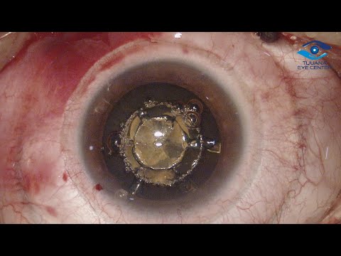


.jpg)
.png)
.png)
.png)


Share this listing