
Comprehensive Cancer Care: Nuclear Medicine in Istanbul Turkey
Modern oncology has evolved far beyond traditional diagnostic imaging and non-targeted interventions. Today, Nuclear Medicine in Istanbul Turkey stands at the absolute forefront of precision oncology, blending molecular biology, advanced radio-chemistry, and state-of-the-art hybrid imaging. Biruni Hospital in Istanbul, Turkey has established itself as an international benchmark for excellence in this field, integrating high-resolution PET/CT technology with specialized radiopharmaceutical production. Through the deliberate application of targeted radioactive materials, nuclear medicine enables oncologists to observe cellular metabolism, map microscopic disease spread, and deliver highly concentrated cellular radiation directly to malignant targets while sparing adjacent healthy tissues.
At Biruni Hospital, the application of radioisotopes is twofold: providing precise diagnostic visualization and offering innovative theranostic treatments. Whether identifying occult metabolic activity through FDG PET/CT, pinpointing specialized receptors using Gallium-68 PSMA and DOTATATE, or revealing subtle tumors via Gallium-68 FAPI, the institution provides comprehensive solutions for complex oncology cases. Furthermore, the hospital's capabilities extend into neuroimaging for cognitive disorders like Alzheimer's and Parkinson's disease, as well as complex infection localization. Supported by an European Association of Nuclear Medicine (EANM) certified theranostics laboratory, Biruni Hospital represents a premier destination for global patients seeking world-class Cancer Care in Istanbul Turkey.
Video Chapters & Quick Navigation
Introduction to Radiopharmaceuticals and Molecular Imaging
Nuclear medicine is a medical specialty that uses small amounts of radioactive materials, known as radiopharmaceuticals or radiotracers, to diagnose, evaluate, and treat various diseases. Unlike structural imaging techniques such as conventional X-rays, Computed Tomography (CT), or Magnetic Resonance Imaging (MRI)—which primarily detail physical anatomy—nuclear medicine focuses on physiological and biochemical function. By tagging biologically active molecules with tiny doses of radioactive isotopes, physicians can trace molecular activity in real time deep within human tissue.
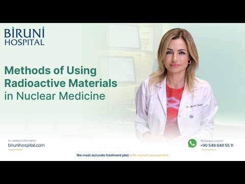
When administered to a patient—typically via intravenous injection, inhalation, or ingestion—these specialized radioactive substances travel to specific organs or cellular receptors. Specialized hybrid cameras, such as Positron Emission Tomography combined with Computed Tomography (PET/CT), detect the gamma rays emitted by the radioisotope. A sophisticated computer system then processes these emissions into three-dimensional tomographic maps of biological function. At [00:00], Biruni Hospital experts emphasize that in clinical practice, radioactive materials serve a dual role: providing high-resolution diagnostic imaging and delivering target-specific radioligand therapy.
The practice of Nuclear Medicine in Istanbul Turkey has advanced dramatically over the past decade. Institutions like Biruni Hospital combine advanced medical physics, radiochemistry, and clinical oncology to transform how complex malignancies are detected, staged, and treated. This functional approach allows medical teams to identify metabolic alterations long before structural anatomical changes become visible on routine anatomical scans, offering patients a crucial lead time in early intervention.
Core Pillars of Modern Nuclear Medicine:
- Molecular Specificity: Utilizing radiolabeled molecules designed to bind exclusively to specific metabolic pathways or cell surface receptors.
- Hybrid Precision: Fusing functional PET images with structural CT scans to establish exact anatomical location alongside physiological activity.
- Theranostic Integration: Transitioning seamlessly from diagnostic imaging to targeted radionuclide therapy using identical targeting vehicles.
- Quantitative Assessment: Measuring standardized uptake values (SUV) to monitor tumor response to therapy with mathematical objectivity.
FDG PET/CT: Mapping Glucose Metabolism in Oncology
The workhorse of molecular imaging across global oncology remains Fluorodeoxyglucose (18F-FDG) PET/CT. FDG is a glucose analog labeled with the radioactive isotope Fluorine-18. Because malignant cells divide rapidly and rely heavily on accelerated glycolysis to sustain their growth—a biological phenomenon known as the Warburg Effect—they absorb significantly higher amounts of glucose compared to healthy surrounding cells. As highlighted at [00:19], FDG PET/CT acts as an essential glucose tracer, allowing nuclear medicine physicians to illuminate cancer cells based on their metabolic demands.
Once injected, FDG enters cells via glucose transporters (GLUT) and undergoes phosphorylation by hexokinase. Unlike normal glucose, however, phosphorylated FDG cannot be fully metabolized in the glycolytic pathway and becomes trapped inside the tumor cell. The radioactive decay of Fluorine-18 emits positrons, which interact with electrons to produce annihilation photons detected by the PET scanner. This mechanism provides exceptional clarity when assessing tumor burdens throughout the body.
FDG PET/CT plays a critical role in comprehensive Cancer Care in Istanbul Turkey across multiple clinical phases. At Biruni Hospital, it is routinely employed for:
- Initial Staging: Determining the primary tumor extent and detecting local or distant lymph node metastases.
- Treatment Response Evaluation: Distinguishing active residual tumor tissue from scar tissue or post-radiation necrosis following chemotherapy or radiotherapy at [00:31].
- Restaging and Recurrence Detection: Locating recurrent disease in patients exhibiting rising tumor markers despite negative findings on standard CT or MRI scans.
- Radiation Therapy Planning: Mapping metabolic tumor volumes to ensure high-dose radiation fields precisely encompass active disease while sparing adjacent critical organs.
By offering safe and consistent monitoring over many years, FDG PET/CT serves as a cornerstone diagnostic protocol for lung cancer, lymphoma, colorectal malignancies, breast cancer, and gynecological cancers at Biruni Hospital.
Targeted Radio-Tracers: Gallium-68 PSMA, DOTATATE, and FAPI
While FDG PET/CT is highly versatile, certain tumors exhibit low glycolytic activity or are situated in organs with high baseline glucose uptake, making FDG less sensitive. To overcome these clinical limitations, nuclear medicine has ushered in second- and third-generation targeted radiotracers labeled with radioisotopes such as Gallium-68 (Ga-68). At [00:43], experts explain how these customized radioactive molecules target specific cell surface receptors with high affinity.
One of the most remarkable breakthroughs in prostate cancer management is Gallium-68 PSMA (Prostate-Specific Membrane Antigen) PET/CT. PSMA is a transmembrane protein overexpressed up to 1,000 times higher on prostate adenocarcinoma cells compared to normal tissue. As discussed at [00:58], Ga-68 PSMA PET/CT enables early detection of primary prostate tumors, precise initial staging, and early identification of biochemical recurrence at extremely low prostate-specific antigen (PSA) blood levels.
For patients diagnosed with neuroendocrine tumors (NETs), Biruni Hospital utilizes Gallium-68 DOTATATE PET/CT. Neuroendocrine tumors frequently express somatostatin receptors (SSTR), particularly subtype 2. As detailed at [01:14], Ga-68 DOTATATE binds selectively to these receptors, allowing nuclear medicine physicians to visualize gastroenteropancreatic neuroendocrine tumors (GEP-NETs), carcinoid tumors, and pheochromocytomas with unparalleled detail and sensitivity.
Furthermore, Biruni Hospital has integrated Gallium-68 FAPI (Fibroblast Activation Protein Inhibitor) PET/CT into its clinical framework at [01:35]. FAPI targets cancer-associated fibroblasts (CAFs), which constitute a major portion of the tumor stroma across more than 28 different cancer types, including gastric, pancreatic, hepatic, and biliary cancers. Because FAPI imaging does not rely on cellular glucose consumption and features low background activity in the brain and liver, it provides exceptional contrast and resolution for hard-to-detect malignancies that are challenging to visualize with conventional FDG imaging.
The EANM Certified Theranostics Laboratory at Biruni Hospital
The synthesis and clinical deployment of specialized radioactive materials require advanced laboratory infrastructure, rigorous quality control, and strict compliance with radiation protection standards. Biruni Hospital features an in-house theranostics radiochemistry laboratory equipped with automated synthesis modules, cleanroom environments, and modern analytical equipment. At [01:19], the facility highlights its dedicated laboratory where advanced radio-tracers are synthesized on-site tailored to immediate patient requirements.
Crucially, this specialized theranostics laboratory has earned official certification from the European Association of Nuclear Medicine (EANM) as noted at [01:28]. EANM certification serves as an international gold standard, validating that the hospital's radiopharmaceutical production, radio-labeling efficiency, purity testing, and patient administration protocols meet European safety and quality standards.
Key Capabilities of Biruni Hospital's EANM Certified Facility:
- In-House Synthesis: On-demand production of short-half-life isotopes including Gallium-68 (Ga-68), Lutetium-177 (Lu-177), and Fluorine-18 (F-18).
- Automated Quality Control: HPLC and TLC testing to verify radiochemical purity exceeding 95% before clinical administration.
- Theranostic Pairing: Smooth transition from diagnostic imaging (e.g., Ga-68 PSMA) to therapeutic intervention (e.g., Lu-177 PSMA radioligand therapy) within the same facility.
- Radiation Safety Compliance: Advanced radiation monitoring systems ensuring complete safety for patients, healthcare workers, and the surrounding environment.
This certified laboratory infrastructure allows Biruni Hospital to deliver high-precision Nuclear Medicine Treatment in Istanbul Turkey, giving international patients seamless access to cutting-edge radioisotope protocols under one roof.
Applications in Neurology, Infection Localization, and Beyond
Beyond its central role in oncological management, PET/CT imaging utilizing radioactive materials provides valuable diagnostic insights across neurology and infectious diseases. At [01:48], clinical specialists highlight that the diagnostic capabilities of PET/CT extend into neurodegenerative disorders and localized inflammatory processes.
The central nervous system relies almost exclusively on glucose for energy metabolism. In neurodegenerative conditions such as Alzheimer's disease, Frontotemporal Dementia, and Parkinson's disease, characteristic patterns of regional glucose hypometabolism emerge long before structural brain atrophy can be detected on standard MRI scans. By conducting FDG PET/CT brain imaging, nuclear medicine specialists at Biruni Hospital can evaluate hypometabolic patterns in the temporoparietal or frontal lobes, aiding in the differential diagnosis of complex dementias at [02:04].
Furthermore, advanced molecular imaging incorporates specific amyloid-targeting radiotracers. Amyloid PET imaging allows clinicians to visualize cortical beta-amyloid plaque deposition in vivo—a key pathological hallmark of Alzheimer's disease. This early, definitive diagnosis enables neurologists to initiate targeted neuro-protective therapies and lifestyle interventions during the early stages of cognitive decline.
Non-Oncology Indications for Molecular Imaging:
- Differential Diagnosis of Dementia: Distinguishing Alzheimer's disease from Lewy body dementia, vascular dementia, or frontotemporal degeneration.
- Refractory Epilepsy Focus Mapping: Identifying interictal hypometabolic zones in candidate patients for surgical resection.
- Fever of Unknown Origin (FUO): Locating hidden infectious or inflammatory foci throughout the body at [02:08].
- Vascular & Prosthetic Infections: Diagnosing aortic graft infections, osteomyelitis, and cardiac device-related endocarditis.
Comparative Overview of Radioactive Tracers in Clinical Practice
Selecting the appropriate radioisotope and molecular carrier is key to maximizing diagnostic sensitivity and treatment precision. The table below outlines the primary radiopharmaceuticals utilized at Biruni Hospital, detailing their target mechanisms, primary clinical applications, and clinical benefits:
| Radioactive Tracer | Biological Mechanism | Primary Clinical Indications | Key Clinical Advantage |
|---|---|---|---|
| 18F-FDG | Glucose metabolic trapping via GLUT transporters and hexokinase activity. | Lymphoma, lung, colorectal, breast cancers; brain metabolic imaging; infection localization. | Universal tumor metabolic indicator; exceptional for monitoring post-treatment response. |
| Gallium-68 PSMA | High-affinity binding to Prostate-Specific Membrane Antigen on cell surface. | Prostate adenocarcinoma staging, biochemical recurrence, theranostic selection. | Detects micrometastases at low PSA levels (<0.2 ng/mL); guides Lu-177 PSMA therapy. |
| Gallium-68 DOTATATE | Selective targeting of Somatostatin Receptor Subtype 2 (SSTR2). | Neuroendocrine tumors (GEP-NETs), carcinoid tumors, pheochromocytomas. | Superior sensitivity over conventional somatostatin receptor scintigraphy (Octreoscan). |
| Gallium-68 FAPI | Targeting Fibroblast Activation Protein (FAP) in tumor microenvironment stroma. | Pancreatic, hepatic, gastric, esophageal cancers, and occult stroma-rich tumors. | High tumor-to-background contrast; independent of glucose metabolism; low brain background. |
The Patient Journey: Experience and Safety at Biruni Hospital
Undergoing nuclear medicine procedures in a foreign country can feel complex for patients and their families. At Biruni Hospital, the clinical pathway is structured to ensure complete patient comfort, minimal wait times, and rigorous safety standards at every stage of diagnosis and treatment.
Upon arrival at Biruni Hospital for Nuclear Medicine Treatment in Istanbul Turkey, patients are welcomed by a multidisciplinary medical team comprising nuclear medicine physicians, medical oncologists, medical physicists, and specialized radiology nurses. The care process follows a carefully coordinated pathway:
- Pre-Procedure Consultation: Clinical assessment, review of prior anatomical imaging (CT/MRI), renal function testing, and verification of fasting or blood glucose protocols.
- Radiopharmaceutical Preparation: In-house synthesis and quality testing of the specific radioisotope (FDG, PSMA, DOTATATE, or FAPI) within the EANM-certified laboratory.
- Precision Injection & Uptake Phase: Intravenous administration of the radiotracer followed by a comfortable, quiet relaxation period (typically 45 to 60 minutes) in dedicated quiet rooms to allow optimal tissue biodistribution.
- Hybrid PET/CT Acquisition: Fast, low-dose image acquisition on high-sensitivity scanners lasting approximately 15 to 20 minutes.
- Multidisciplinary Reporting: Joint review of reconstructed 3D fusion images by nuclear medicine specialists and consulting oncologists to formulate actionable treatment recommendations.
Patient safety is maintained at all times. The radiation exposure from diagnostic PET/CT scans is low, comparable to standard multi-slice diagnostic CT procedures. Radioisotopes decay rapidly and are eliminated naturally from the body through hydration and renal excretion, ensuring minimal radiation exposure for both patients and their families.
The Future of Nuclear Medicine and Theranostics in Turkey
The field of nuclear medicine is undergoing a transformative shift toward theranostics—a term derived from the combination of therapeutics and diagnostics. Theranostics embodies the ultimate vision of personalized medicine: "we see what we treat, and we treat what we see." By using one radiopharmaceutical to image and locate cancer receptors, clinicians can swap the diagnostic isotope for a therapeutic, beta- or alpha-emitting radioisotope (such as Lutetium-177 or Actinium-225) to destroy targeted tumor cells with pinpoint accuracy.
As a leading center for Cancer Care in Istanbul Turkey, Biruni Hospital continues to expand its theranostic initiatives. The clinical integration of Lutetium-177 PSMA therapy for metastatic castration-resistant prostate cancer (mCRPC) and Lutetium-177 DOTATATE peptide receptor radionuclide therapy (PRRT) for advanced neuroendocrine tumors offers new options for patients who have exhausted traditional systemic therapies.
With ongoing research into targeted alpha therapies (TAT), novel FAPI-based therapeutic combinations, and artificial intelligence-assisted image quantification, Biruni Hospital remains dedicated to advancing nuclear medicine. By bridging modern molecular science with compassionate patient care, the institution provides new hope and improved clinical outcomes for oncology patients worldwide.
Ready to Explore Modern Cancer Care at Biruni Hospital?
PlacidWay Medical Tourism connects you with top-rated Cancer Care in Istanbul Turkey and oncology centers at Biruni Hospital. Access advanced nuclear medicine imaging, certified theranostic treatments, and personalized oncology care tailored to your health needs.
GET A FREE QUOTEView Full Video Transcript
[00:00:00] In nuclear medicine, we use radioactive materials for both imaging and treatment.
[00:00:01] We perform both imaging and treatment
[00:00:03] using radioactive materials. Let's talk about PET/CT,
[00:00:05] which is the most frequently used and also the best-known method among the public.
[00:00:07] In PET/CT,
[00:00:09] we use different radioactive materials
[00:00:11] for the diagnosis of various cancers
[00:00:13] and for evaluating treatment response,
[00:00:15] and we have been safely monitoring
[00:00:17] our patients for many years.
[00:00:19] Among these, the best known
[00:00:21] is FDG PET/CT, which is a glucose analog.
[00:00:24] We administer this glucose analog to our patients
[00:00:27] after labeling it with a radioactive substance,
[00:00:29] allowing us to visualize cancer cells.
[00:00:31] For diagnosis, evaluating the treatment response
[00:00:33] of patients who have received chemotherapy or radiotherapy,
[00:00:35] and detecting distant organ metastases
[00:00:36] in patients who have undergone surgery,
[00:00:38] we successfully use PET/CT imaging
[00:00:41] in clinical practice.
[00:00:43] However, it is not only FDG PET/CT;
[00:00:46] with current research and developments in nuclear medicine,
[00:00:48] we also use different radioactive materials
[00:00:50] for the diagnosis and imaging
[00:00:52] of different types of tumors,
[00:00:54] and we have other radioactive agents
[00:00:55] that we successfully use for these purposes.
[00:00:57] One of the best known
[00:00:58] is imaging with Gallium-68 PSMA PET/CT.
[00:01:02] In patients diagnosed with prostate cancer,
[00:01:04] Gallium-68 PSMA PET/CT imaging
[00:01:06] plays an effective role in both
[00:01:09] the diagnosis and treatment of our patients.
[00:01:11] In addition to Gallium-68 PSMA PET/CT,
[00:01:14] we also successfully use Gallium-68 DOTATATE PET/CT
[00:01:18] in patients diagnosed with neuroendocrine tumors.
[00:01:19] We have a special theranostics laboratory
[00:01:21] where these radioactive materials
[00:01:23] are specially prepared at our hospital.
[00:01:25] This laboratory has also received certification
[00:01:28] from the European Association of Nuclear Medicine,
[00:01:30] and we carry out these productions
[00:01:32] in our laboratory.
[00:01:33] In addition, beyond these agents,
[00:01:35] with Gallium-68 FAPI PET/CT, which was introduced into use
[00:01:38] relatively recently,
[00:01:41] we can also image tumors that we have not been able to visualize
[00:01:43] with these other agents,
[00:01:45] with high accuracy and resolution.
[00:01:48] In addition to imaging cancer patients,
[00:01:49] we also successfully use PET/CT
[00:01:52] for other diseases.
[00:01:54] The area in which we use it most frequently
[00:01:56] is brain imaging. In Alzheimer's disease
[00:01:58] or Parkinson's disease,
[00:02:02] in addition to FDG PET/CT,
[00:02:02] when we perform amyloid imaging with PET/CT using other agents,
[00:02:04] we can also obtain successful imaging results
[00:02:06] in the differential diagnosis of dementia in our patients.
[00:02:08] We can also use PET/CT from time to time
[00:02:10] for infection imaging.
[00:02:16] [Music]



















Share this listing