It is incredibly stressful when a family member undergoes a medical procedure, only to find the results falling short of expectations. Many patients and their families face this exact dilemma when searching for the root causes of blurred vision after initial eye procedures. Advanced corrective cataract surgery in India has emerged as a highly reliable solution for addressing these unexpected and complex surgical complications.
When a standard lens replacement fails to restore clear sight, an immediate and thorough clinical evaluation is mandatory to prevent long-term damage. Specialized ophthalmologists utilize state-of-the-art diagnostic imaging to pinpoint mechanical failures within the eye. Identifying issues such as a displaced intraocular lens allows surgeons to execute rapid, minimally invasive corrections.
Modern advancements in ophthalmology mean that even complex post-operative problems often have straightforward, fast solutions. Patients experiencing poor vision after lens replacement do not have to live with permanent impairment. By consulting with experts in secondary eye interventions, individuals can fully restore their visual acuity and regain their quality of life.
Video Chapters
Common Causes of Blurred Vision After Initial Eye Procedures
Experiencing cloudy or obstructed sight following an eye operation is a major source of anxiety for patients worldwide. Many individuals immediately assume that the operation was completely botched or that permanent damage has occurred. In reality, temporary blurring can be a standard part of the healing process, though persistent issues require immediate medical investigation.
One frequent complication is corneal edema, which involves the swelling of the eye's clear front surface. This swelling typically subsides within a few days when treated with prescribed anti-inflammatory drops. However, if the blurriness remains constant or worsens, it signals a deeper mechanical or biological problem that needs expert attention.
Another prevalent issue is posterior capsule opacification (PCO), often referred to as a secondary cataract. PCO occurs when cellular growth clouds the thin membrane holding the new artificial lens in place. While PCO is easily treated with a quick laser procedure, other complications involve the physical positioning of the lens implant itself.
The Impact of Surgical Execution
The success of phacoemulsification relies heavily on the precise execution of each surgical step. If the native lens fragments are not entirely removed, retained material can cause severe intraocular inflammation. This localized inflammation directly contributes to elevated eye pressure and significant visual distortion.
Furthermore, the architectural integrity of the surgical incisions plays a crucial role in post-operative sight. Poorly constructed incisions can induce astigmatism, altering how light refracts onto the retina. Correcting these surgically induced astigmatisms often requires a secondary intervention by an expert ophthalmologist.
Understanding Intraocular Lens (IOL) Anatomy and Surgical Failures
To comprehend why complications occur, one must understand the basic anatomy of an artificial intraocular lens. Modern IOLs are predominantly composed of two main structural elements: the optic and the haptics. The optic is the central, transparent circular zone responsible for refracting light and focusing images.
The haptics are the flexible, arm-like extensions attached to the periphery of the optic. These structures are designed to anchor the lens securely within the eye's natural capsular bag. Proper deployment of the haptics is absolutely essential to keep the optic perfectly centered along the patient's visual axis.
During surgery, these lenses are tightly folded and inserted through a microscopic incision using a specialized injector. Once inside the eye, the lens should naturally unfold and settle into its permanent position. However, as noted in the clinical case study at [00:30], there are instances where the haptic completely fails to separate from the optic.
Why Lenses Fail to Unfold
Several technical factors can prevent an artificial lens from deploying correctly inside the eye. The temperature of the operating room, the specific material of the lens, and the viscosity of protective gels used during surgery all play a role. Hydrophobic acrylic lenses, for instance, are highly temperature-sensitive and may remain stiff if the environment is too cold.
Additionally, if the protective viscoelastic gel is not adequately flushed from the eye, it can trap the haptic against the optic body. This mechanical adhesion prevents the lens from centering, causing it to sit entirely outside the patient's line of sight. This precise malfunction results in the severe visual impairment described by the distressed patient.
Diagnosing Displaced Lenses in Post-Operative Eye Care
When a patient presents with unexpectedly poor vision after a routine procedure, a meticulous diagnostic protocol must be initiated. A standard visual acuity test is insufficient for identifying complex anatomical failures within the anterior chamber. Surgeons must rely on advanced slit-lamp biomicroscopy to examine the microscopic structures of the eye.
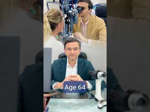
In the highlighted scenario of the 64-year-old male [00:15], the initial surgery was performed correctly in terms of cataract removal. However, a detailed post-operative evaluation revealed that the artificial lens was completely missing from the central optical zone. The lens had remained bundled and stuck to itself, effectively leaving the eye without a focusing mechanism.
Comprehensive pupillary dilation is required to visualize the peripheral edges of the capsular bag where the haptics rest. Without dilation, a stuck haptic hidden behind the iris might be completely overlooked by an inexperienced practitioner. This emphasizes why seeking an expert second opinion is vital for resolving unexplained blurry vision after lens replacement.
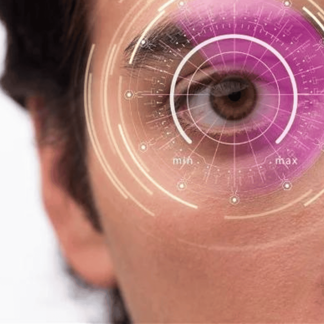
The Importance of Advanced Imaging
Modern clinics often employ optical coherence tomography (OCT) to map the exact location of the misaligned lens. High-definition OCT scans provide a cross-sectional view of the eye, revealing the exact degree of haptic adhesion. This imaging allows the surgeon to formulate a precise surgical plan before returning the patient to the operating room.
Diagnostic ultrasound is another vital tool utilized when corneal swelling obscures direct visual inspection of the lens. These rigorous diagnostic steps ensure that the subsequent corrective procedure is targeted, safe, and effective. Accurate diagnosis is the cornerstone of successful secondary eye interventions.
The Five-Minute Corrective Procedure for Dislocated Intraocular Lenses
Once a stuck haptic is diagnosed, the solution is highly surgical but surprisingly swift for the patient. The individual is prepped and returned to the sterile environment of the operating theater. As mentioned in the clinical overview at [00:45], resolving this complex mechanical problem often takes merely five minutes.
Under local or topical anesthesia, the surgeon re-enters the eye through the original microscopic incisions. Using specialized micro-instruments, such as a Sinskey hook, the surgeon carefully navigates to the trapped lens. Gentle, precise physical manipulation is applied to separate the adhered haptic arm from the central optic.
As soon as the haptic is released, the intrinsic memory of the lens material causes it to spring into its proper shape. The lens immediately centers itself within the capsular bag, falling perfectly into alignment with the visual axis. The surgeon then meticulously removes any remaining protective gels before sealing the tiny incisions.
Immediate Post-Operative Results
The visual recovery following this specific corrective intervention is often incredibly dramatic and immediate. By the very next morning, patients typically experience a profound restoration of visual clarity. The frustration and anxiety of a failed initial surgery are instantly alleviated.
This rapid turnaround demonstrates why complex post-operative visual problems often have very simple solutions. It reinforces the necessity of skilled surgical intervention rather than adopting a "wait and see" approach for poor vision. Prompt action protects the eye and guarantees the intended outcome of the original procedure.
Evaluating Advanced Intraocular Lens Options for Superior Vision
Choosing the correct intraocular lens prior to surgery dramatically influences the long-term visual outcome. Patients have access to a variety of sophisticated lens technologies designed to match their specific lifestyle requirements. Understanding these options helps set realistic expectations for post-operative visual acuity.
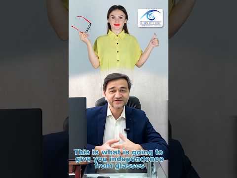
Standard monofocal lenses provide excellent clarity at a single distance, usually optimized for driving and watching television. However, patients opting for monofocal lenses will still require reading glasses for close-up tasks. For those seeking greater independence from eyewear, premium lens options offer advanced refractive capabilities.
Multifocal and Toric lenses are highly engineered implants that correct astigmatism and provide a wider range of focus. These complex lenses require absolute precision in alignment during the surgical procedure. If a premium lens suffers from a stuck haptic, the visual distortion is notably more severe than with standard lenses.
Comparison of IOL Technologies
| Lens Type | Primary Function | Best Suited For | Surgical Considerations |
|---|---|---|---|
| Monofocal | Provides clear vision at one set distance. | Patients comfortable wearing reading glasses. | Most common, highly stable inside the eye. |
| Toric | Corrects pre-existing corneal astigmatism. | Patients with uneven corneal curvature. | Requires exact rotational alignment during surgery. |
| Multifocal | Splits light for near, intermediate, and far vision. | Individuals wanting total independence from glasses. | Highly sensitive to slight displacements or stuck haptics. |
| EDOF | Extended Depth of Focus for a continuous visual range. | Active lifestyles requiring good intermediate vision (computers). | Less glare at night compared to traditional multifocals. |
Essential Post-Operative Recovery Protocols for Secondary Eye Interventions
Undergoing a secondary corrective eye surgery requires strict adherence to post-operative care guidelines to ensure optimal healing. Although the procedure to release a stuck haptic is rapid, the eye has still endured physical manipulation. Protecting the delicate ocular structures is paramount in the days immediately following the intervention.
A rigorous regimen of pharmacological eye drops is prescribed to prevent infection and control internal inflammation. Broad-spectrum antibiotic drops mitigate the risk of bacterial endophthalmitis, a severe post-surgical complication. Simultaneously, topical corticosteroids are applied to soothe the eye tissues and promote swift cellular recovery.
Patients are explicitly instructed to avoid rubbing or applying any external pressure to the treated eye. Engaging in strenuous physical activities, lifting heavy objects, or bending over can dangerous spike intraocular pressure. Following these behavioral restrictions guarantees that the newly positioned intraocular lens remains perfectly stable.
Key Recovery Steps to Follow
- Consistent Medication Application: Administer all antibiotic and anti-inflammatory eye drops strictly according to the surgeon's schedule.
- Protective Eyewear: Wear a protective ocular shield while sleeping to prevent accidental rubbing or trauma during the night.
- Water Avoidance: Keep non-sterile tap water, soap, and shampoo out of the eye for at least one week to prevent chemical irritation and infection.
- Follow-Up Compliance: Attend all scheduled post-operative evaluations so the ophthalmologist can monitor internal healing and lens stability.
Why Medical Tourists Choose Corrective Cataract Surgery in India
India has rapidly ascended as a premier global destination for advanced ophthalmological procedures and corrective eye surgeries. The country boasts a vast network of internationally accredited medical facilities equipped with the latest diagnostic and surgical technologies. Patients suffering from unresolved visual complications often travel to major hubs like Mumbai to seek out elite medical expertise.
One of the primary drivers of this medical tourism boom is the presence of highly specialized, globally trained eye surgeons. These professionals handle an immense volume of complex cases, granting them unparalleled experience in managing intraocular lens complications. This high case volume translates directly into superior surgical outcomes and innovative problem-solving in the operating room.
Beyond surgical excellence, the cost-effectiveness of comprehensive eye treatments in India is incredibly attractive to international patients. The cost of complex corrective eye surgery in Mumbai is a fraction of the price charged in North America or Europe. This significant financial advantage is achieved without any compromise to patient safety, clinical hygiene, or the quality of premium lens implants used.
Advantages of the Indian Healthcare Ecosystem
International patients benefit immensely from the zero wait-time policies prevalent in premier Indian eye clinics. Unlike state-funded healthcare systems in the West, private clinics in India can schedule urgent secondary interventions within days. Rapid treatment is essential when dealing with incorrectly positioned lenses that are causing debilitating vision loss.
Furthermore, English is the primary language of communication in the Indian medical sector, eliminating stressful language barriers for foreign travelers. Clinics often provide dedicated international patient coordinators who manage travel logistics, medical visas, and local accommodations. This holistic approach to patient care makes the journey for complex restorative surgery virtually seamless.
Selecting the Best Ophthalmologist for Complex IOL Repositioning
Choosing the right specialist to correct a botched or complicated initial eye surgery is a critical decision. Not all ophthalmologists possess the micro-surgical dexterity required to safely manipulate a folded lens within a delicate capsular bag. Patients must actively seek out surgeons who specialize specifically in secondary interventions and complex anterior segment reconstructions.
Evaluating a surgeon's credentials involves reviewing their fellowship training, clinical affiliations, and specific experience with unreleased haptics. A premier surgeon will heavily rely on transparent patient communication, thoroughly explaining the exact mechanical failure causing the blurry vision. They will provide a clear, step-by-step roadmap detailing exactly how the five-minute corrective procedure will resolve the issue.
Ultimately, restoring visual acuity after a surgical disappointment requires profound trust between the patient and the medical team. Clinics equipped with advanced surgical microscopes and proven track records offer the highest probability of visual success. By prioritizing technical expertise and advanced diagnostic capabilities, patients ensure their corrective cataract surgery is safe, effective, and permanent.
Struggling with Poor Vision After Eye Surgery?
Do not let surgical complications compromise your quality of life. Connect with elite ophthalmologists in India for a comprehensive evaluation and discover affordable, expert solutions for permanent visual clarity.
Get Free QuoteView Video Transcript
[00:00] Parent ki cataract surgery hui par phir bhi unhe nahi dikh raha.
[00:05] Soche aapke parent hai jiski age 64 hai. And aap yeh dilemma mein hai ki itni vyavasthit surgery hone ke baad bhi unko dikh kyun nahi raha.
[00:15] Aisa hi ek case mere paas aaya tha. 64 year old male, jiski surgery hui thi paas mein hi kahin aur.
[00:20] Aur jab maine unko dekha to I was shocked to see ki lens uski jagah pe nahi tha. Halanki surgery was absolutely fine.
[00:30] On detailed evaluation mujhe yeh pata chala ki lens ki jo optic hoti hai jisse hum dekhte hai, aur haptic hoti hai jisse lens supported rehta hai, woh chipke hue the. Aur release nahi kiya gaya tha.
[00:40] Solution bahut hi simple tha. Unko surgery ke liye liya.
[00:45] Hardly 5 minute ki procedure mein dono ko release kiya. Aur next day itself patient ko ekdam vyavasthit dikhta hai.
[00:55] Complex lagti hai baatein par hoti bahut simple hai solutions.


.jpg)
.png)
.png)
.png)


Share this listing