
Undergoing a complete dental restoration is a significant personal and clinical milestone. This visual journey captures the reality of a modern dental treatment process, illustrating what a patient experiences from their initial clinic arrival to the final mirror reveal.
Quick Takeaway: A comprehensive smile makeover follows a highly structured clinical pathway: it begins with an initial medical intake and panoramic diagnostic imaging, advances through digital treatment planning, and concludes with the precise clinical preparation and bonding of the final restorations.
Establishing the Baseline: Arrival and Medical Intake
When a patient decides to pursue a major dental transformation, the journey starts long before any clinical work begins. The opening moments of the video show the patient arriving at the clinic and reviewing intake documents with the staff. While this may seem like routine administrative work, the medical and dental intake is a foundational safety requirement for any major procedure.
For patients undergoing multiple restorations, crowns, or potential implant placements, the clinical team must establish a comprehensive health baseline. This involves disclosing current medications, underlying medical conditions, and any history of allergic reactions to local anesthetics. Because oral health is intimately connected to systemic health, seemingly unrelated conditions—such as cardiovascular issues or autoimmune disorders—can dictate the type of anesthetics used or the required healing timelines.
Furthermore, this initial stage is when the logistical framework of the treatment is finalized. For those traveling as part of a medical tourism journey, this step often includes reviewing the treatment timeline to ensure all clinical phases can be completed within the patient's travel window. A thorough intake process is the first indicator that a clinic prioritizes patient safety alongside aesthetic outcomes, setting an organized tone for the subsequent clinical phases.
Seeing Beneath the Surface: The Role of Diagnostic Imaging
Visual examinations only reveal a fraction of a patient’s actual oral health. Early in the footage, the patient is shown being positioned in a panoramic X-ray machine. This technology captures a two-dimensional flat image of the entire mouth, including the jaws, teeth, sinuses, and surrounding bone structure in a single continuous scan.
Before any aesthetic changes can be planned, the restorative dentist must verify that the biological foundation is healthy. Placing permanent porcelain restorations on teeth with hidden decay, infected roots, or compromised bone support will inevitably lead to structural failure and complex complications down the road. The diagnostic imaging phase allows the clinical team to identify issues that are invisible to the naked eye.
- Evaluating Bone Density: Ensures that there is sufficient foundational support, which is especially crucial if dental implants are part of the restorative plan.
- Identifying Hidden Decay: Confirms that the natural teeth are biologically healthy enough to securely hold permanent veneers or crowns without future failure.
- Mapping Nerve Pathways: Provides an exact map of the oral anatomy, allowing the dentist to prevent nerve damage during any surgical or aggressive preparative phases.
If a patient’s treatment plan is particularly complex, imaging becomes even more critical. While a standard panoramic X-ray provides an excellent overview, advanced cases often require a 3D Cone Beam Computed Tomography (CBCT) scan. By relying on precise diagnostic data, the dental team can transition from the evaluation phase to the design phase with complete confidence in the underlying oral structure.
Designing the Result: Digital Treatment Planning and Consultation
The days of unpredictable dental outcomes have largely been replaced by digital precision. In the video, the dentist and patient are seen reviewing documents and digital screens together. This collaborative consultation is arguably the most critical decision-making point in the entire smile makeover process.
Modern CAD/CAM (Computer-Aided Design and Computer-Aided Manufacturing) technologies allow providers to model the final outcome before a single tooth is touched. During this phase, the dentist evaluates the patient’s facial symmetry, lip line, and gum architecture. The goal is to design restorations that look natural and function harmoniously with the patient's unique facial structure.
Mapping the exact oral anatomy and clearly documenting the patient's functional and aesthetic goals.
Creating a visual or physical prototype of the final teeth to align expectations before any enamel is altered.
Removing the necessary tooth structure and placing temporary restorations while the final ceramics are crafted.
Permanently bonding the veneers or crowns and meticulously checking the bite to ensure long-term structural safety.
This planning phase is also where patient expectations are aligned with clinical realities. The patient and provider must agree on the shade (color), shape, and length of the new teeth. While a patient may desire the brightest possible shade, the dentist will offer guidance on what will look most natural based on the patient's complexion. The digital mock-up acts as a blueprint, ensuring that the patient fully understands the proposed changes and eliminating surprises on the final day of treatment.
The Clinical Phase: Tooth Preparation and Restorative Work
The actual physical transformation takes place in the treatment chair. The footage highlights this clinical phase, showing the dentist actively working on the patient's teeth. Whether the treatment plan calls for porcelain veneers or full-coverage crowns, the natural teeth must be prepared to receive the new restorations.
Preparation involves carefully removing a specific amount of the tooth's outer enamel layer. This step is necessary to create space for the ceramic material so that the final teeth do not appear bulky or artificial. For standard veneers, this reduction might be minimal. For dental crowns, a more significant portion of the tooth structure must be reduced to allow the crown to encase the entire tooth securely.
| Restoration Type | Clinical & Practical Reality |
|---|---|
| Porcelain Veneers | Primarily addresses aesthetic concerns on the front surface of the teeth. Requires minimal enamel removal, but remains an irreversible procedure. |
| Full-Coverage Crowns | Restores severely damaged, weakened, or decayed teeth. Encases the entire tooth structure, requiring more significant preparative reduction for stability. |
It is vital for patients to understand that this preparative step is generally irreversible. Once enamel is removed, the tooth will always require a restoration. Because this can be an invasive process, local anesthesia is administered to ensure the patient remains completely comfortable. After the teeth are prepared, the dentist captures the new shape—either using traditional putty or a high-speed digital intraoral scanner. These precise dimensions are sent to a specialized dental laboratory where the permanent ceramics are custom-fabricated. In the interim, the patient is fitted with temporary restorations that protect the prepared teeth.
The Final Reveal: Evaluating the New Smile and Bite Mechanics
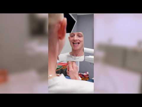
The culmination of the smile makeover journey is the final fitting and cementation, highlighted in the video by the patient examining his new smile in a hand mirror. This moment is often highly emotional, reflecting the profound psychological impact of a restored smile. However, from a clinical perspective, this step involves rigorous mechanical testing before the restorations are permanently bonded.
Before the final curing light is applied, the dentist places the veneers or crowns on the teeth using a temporary try-in paste. This allows both the patient and the dentist to evaluate the aesthetics, fit, and color in natural light. If any final adjustments are needed, they can be made seamlessly at this stage.
"The final mirror check represents more than just a cosmetic change—it marks the restoration of functional confidence and the culmination of a highly precise clinical journey."
Once the aesthetics are approved, the teeth are meticulously cleaned and etched, and the restorations are permanently bonded into place. The dentist then spends a significant amount of time checking the patient's occlusion (bite). The upper and lower teeth must meet perfectly; even a fraction of a millimeter of high contact can cause jaw pain, muscle fatigue, or catastrophic chipping of the new porcelain. The enthusiastic reaction of the patient in the footage underscores the success of this careful alignment, resulting in a smile that is structurally sound and visually transformative.
Protecting the Investment: Long-Term Maintenance of Dental Restorations
A newly restored smile is a major investment in one's health and confidence, but it is not entirely maintenance-free. While porcelain and ceramic materials will not decay, the natural tooth structure underneath and the surrounding gum tissue remain fully susceptible to bacterial plaque, cavities, and periodontal disease.
Patients must commit to a rigorous daily oral hygiene routine. This includes brushing with a non-abrasive toothpaste to avoid scratching the polished ceramic surface, and diligent flossing to maintain the health of the gums around the margins of the restorations. Unhealthy gums can recede, exposing the natural root of the tooth and compromising the aesthetic line of the veneer or crown.
Before committing to a smile makeover, ask your provider about the expected lifespan of the materials used, the specific daily maintenance required, and whether a protective night guard will be necessary to prevent the restorations from chipping during sleep.
Additionally, the mechanical forces of the human jaw are immensely powerful. Patients who suffer from bruxism (teeth grinding or clenching), particularly at night, run a high risk of fracturing their new restorations. Providers routinely recommend a custom-fitted acrylic night guard to protect the investment while the patient sleeps. Regular professional cleanings and check-ups remain essential to monitor the integrity of the bonding, evaluate gum health, and ensure the long-term success of the dental work.
Discuss Your Dental Restoration Options With a Specialist
Understanding the steps, materials, and clinical requirements for a smile makeover is essential before making a decision. Connect with a qualified dental provider to review your specific oral health needs, explore suitable treatment options, and plan your care effectively.
Request a Dental ConsultationView Video Transcript
[Note: This video features a visual journey set to background music with no spoken dialogue. The following describes the on-screen events.]
[0:00] - The patient arrives in the clinic lobby and reviews intake paperwork with the staff.
[0:09] - The patient is positioned inside a panoramic X-ray machine for diagnostic imaging.
[0:15] - The clinic team reviews a detailed treatment plan document and digital imaging.
[0:17] - The dentist sits with the patient in the treatment room for a pre-procedure consultation.
[0:22] - The dentist performs a clinical examination and prepares the teeth for restoration.
[0:27] - The dentist shows the patient digital references of the treatment on a smartphone.
[0:31] - A dental assistant helps prepare the patient for the final bonding phase.
[0:34] - The patient looks into a hand mirror, evaluating the final restored teeth, and smiles broadly.
[0:42] - The patient continues to admire the final results.




























Share this listing