Advanced Guided Dental Implantation Procedure in Hungary
If you are exploring advanced options for tooth replacement, understanding the guided dental implantation procedure in Hungary is an essential first step. This innovative clinical approach offers a highly accurate alternative to traditional freehand methods, transforming how oral surgeons execute complex full mouth restorations. By leveraging state-of-the-art diagnostic technology and digital software, patients experience significantly fewer complications and highly predictable structural outcomes.
Thousands of individuals travel abroad annually to access world-class dental care at competitive price points, making the surgical tourism sector for computer-guided implants increasingly popular. This advanced technique specifically minimizes surgical soft tissue trauma, improves titanium root placement accuracy, and drastically accelerates the overall postoperative recovery timeline. In the detailed sections below, we analyze the intricate mechanical details of guided implant surgery and outline why it represents the undisputed gold standard in modern oral rehabilitation.
Video Chapters & Quick Navigation
- Guided Implants & Biological Mechanisms
- Guided Surgery vs Freehand Implantation
- Digital Planning for Full Mouth Restoration
- Utilizing CT Scans for Exact Placement
- The Role of 3D Printed Surgical Templates
- Flapless Dental Implant Surgery Trauma
- Accelerating Surgery Recovery Time
- Choosing the Best Dental Specialist
Understanding Guided Dental Implants and Their Biological Mechanisms
Guided dental implants involve using advanced computer software to virtually map the exact location of a titanium root before any actual surgery takes place. This strictly digital approach removes the spatial guesswork historically associated with older surgical methods. The surgeon evaluates the software to identify the densest bone areas, ensuring the implant achieves maximum primary stability immediately upon insertion.
Avoiding critical anatomical structures is a major component of this digital mapping protocol. The exact distance between the proposed implant tip and the mandibular nerve is measured down to the millimeter. As noted during the clinical overview, a guided implantation technique definitively precludes inaccuracy compared to standard medical freehand placement [00:18]. The predetermined mechanical path guarantees absolute patient safety during the high-speed drilling sequence.
The Biological Need for Primary Stability
The biological mechanisms of cellular osseointegration depend heavily on this initial surgical stability. When a titanium implant is placed firmly into dense bone without micromovements, surrounding osteoblast cells adhere rapidly to the textured metal surface. A proper computer-guided dental implant surgery protocol ensures the fixture is driven into the bone at the exact angle required to distribute daily chewing forces evenly.
Proper angulation dictates the structural success of the final prosthetic ceramic crown. Implants placed at awkward spatial angles require highly customized abutments that can severely compromise the mechanical integrity of the artificial tooth. Digital surgical guidance ensures the implant aligns perfectly with the patient's natural bite plane, dramatically simplifying the laboratory restorative phase.
Guided Implant Surgery vs Medical Freehand Implantation
For several decades, medical freehand implantation served as the only clinical option available for replacing missing roots. Dental surgeons relied entirely on 2D periapical x-rays, visual tissue inspection, and their own tactile experience to estimate bone depth. While highly skilled practitioners routinely achieved excellent success rates, the lack of a physical surgical guide always introduced a variable margin of spatial deviation.
Comparing guided vs freehand dental implant placement highlights a massive technological leap forward in modern restorative dentistry. A purely freehand approach can occasionally result in accidental perforation of the delicate sinus cavity if the drill angle shifts slightly. In stark contrast, computer-guided surgery utilizes a rigid mechanical stop that physically prevents the drill from advancing past the safe, pre-calculated millimeter depth.
Many patients demand to know exactly how these two distinct methods differ regarding physical surgical trauma and procedural speed. The comparative table below outlines the specific clinical characteristics separating guided digital protocols from traditional manual implant placement.
| Feature | Guided Implant Surgery | Medical Freehand Implantation |
|---|---|---|
| Incision Requirement | Flapless (small tissue punch) | Large scalpel incisions necessary |
| Surgical Accuracy | Sub-millimeter mechanical precision | Dependent on visual spatial estimation |
| Drill Depth Control | Mechanical hard-stops on the template | Manual depth gauge observation |
| Procedure Speed | Significantly faster clinical chair time | Longer due to continuous manual verification |
Step-by-Step Digital Planning Process for Full Mouth Restoration
The foundation of any successful surgical outcome is established extensively before the actual operation day. Every individual case begins with a rigorous planning process alongside a baseline condition survey [00:33]. This initial survey clinically assesses periodontal health, occlusal wear patterns, and the presence of any active dental bone infections.
Digital planning for full mouth dental implants utilizes specialized 3D clinical software to merge various patient data points. The clinical laboratory team imports optical digital impressions of the patient's gums into the central program. This precise data layer allows the restorative dentist to design a highly accurate virtual wax-up of the final prosthetic teeth before surgery begins.
Top-Down Treatment Planning Methods
Working backward logically from the final tooth position is known in the industry as top-down treatment planning. Once the ideal ceramic crown position is finalized, the surgeon places the virtual titanium implant directly underneath it within the software environment. This synchronization ensures the surgical and restorative phases align flawlessly, completely eliminating compromised aesthetics later on.
During this specialized phase, jaw bone density is analyzed layer by microscopic layer. If the software detects critically insufficient bone volume in the posterior maxilla, the surgeon preemptively schedules a sinus lift bone grafting procedure. This level of digital foresight prevents intraoperative complications that routinely delay standard traditional treatments.
Utilizing CT Scans and Panoramic X-Rays for Exact Placement
Advanced diagnostic imaging is completely non-negotiable for executing a computer-guided oral surgery. Standard 2D periapical x-rays simply cannot provide the necessary volumetric spatial data to execute a flawless surgical plan. The precise spatial location and proper size of the implants are exclusively determined by evaluating the CT scan, the panoramic x-ray, and a physical study sample [00:41].
The panoramic x-ray supplements the raw 3D data by providing a flat, comprehensive global view of the entire jawline geometry. This broad image is excellent for identifying widespread bone loss patterns or impacted wisdom teeth that might interfere with the anterior surgical field. Cross-referencing the wide panoramic view with the localized volumetric data guarantees absolute diagnostic accuracy.
Cone Beam Computed Tomography (CBCT) Imaging
A Cone Beam Computed Tomography (CBCT) scan for accurate dental implant placement acts as the master roadmap for the entire procedure. It captures a 360-degree view of the maxillofacial skeleton, accurately rendering soft tissues, nerve canals, and bone density in high definition. Surgeons utilize the precise Hounsfield units derived from the scan to measure bone hardness, which directly dictates the specific drilling speed required.
The physical study sample, which is typically captured via an intraoral optical scan, digitally records the exact topography of the gingival tissues. Merging this soft surface scan with the underlying rigid skeletal CBCT data creates a perfect dual-layered digital clone of the patient. This sophisticated digital clone is exactly what the software uses to calculate the final drill trajectories.
The Essential Role of 3D Printed Surgical Templates in Modern Clinics
Translating the virtual surgical plan into a physical clinical reality requires specialized laboratory manufacturing equipment. Following the software design phase, the clinic automatically creates a custom drilling template utilizing an advanced 3D printer [00:49]. This device prints a highly accurate, biocompatible surgical resin guide that perfectly matches the exact contours of the patient's oral cavity.
A 3D printed surgical guide for dental implants acts as a rigid, unmoving stencil during the surgical operation. It snaps securely onto the adjacent anchor teeth or rests flush against the edentulous mucosal tissue, locking the pre-planned drill paths rigidly into place. Modern in-house 3D printing has completely revolutionized the turnaround time for complex implant treatments globally.
Integrating Titanium Sleeves for Drill Accuracy
Embedded firmly within the printed resin guide are medical-grade titanium sleeves that correspond mathematically to the specific diameter of the surgical drills. These metal sleeves serve multiple critical mechanical functions during the bone osteotomy phase. They strictly prevent the high-speed drill bit from vibrating or drifting off-center while penetrating the dense cortical bone layers.
Furthermore, these titanium sleeves guide the sterile cooling saline irrigation directly into the active drill site seamlessly. This continuous liquid cooling process prevents thermal necrosis of the surrounding living bone cells. Maintaining bone vitality is absolutely crucial for ensuring the titanium threads integrate without rejection.
Flapless Dental Implant Surgery: Minimizing Tissue Trauma and Cutting
The most noticeable patient benefit derived from using a 3D surgical template is the drastic reduction in physical soft tissue trauma. Leveraging this exact spatial technique, highly complex procedures can be executed perfectly without traditional tissue cutting and subsequent lobe formation [00:56]. This streamlined surgical method is universally recognized globally as flapless dental implant surgery.
Standard oral surgery protocols historically mandate using a sharp scalpel to slice the gingiva and forcefully peel back a soft tissue flap. This gross reflection unnecessarily exposes the raw alveolar bone to the oral environment and severs thousands of microscopic blood vessels. Advanced flapless dental implant surgery techniques bypass this highly invasive step entirely by utilizing a precise, small circular tissue punch.
Preserving the Periosteum Blood Supply
The specialized tissue punch removes only the exact millimeter of gum tissue strictly necessary to insert the titanium root cylinder. Because the periosteum—the delicate connective tissue layer covering the bone—remains entirely untouched, the vital blood supply to the surgical site is perfectly preserved. Intact robust vascularization aggressively supports the immediate cellular repair response needed for successful osseointegration.
Avoiding large scalpel incisions completely eliminates the need for complex, uncomfortable suturing at the end of the clinical appointment. Patients depart the dental clinic without painful stitches pulling tightly on their inflamed gums. The structural integrity of the natural gum line remains structurally intact, ensuring a vastly more natural-looking emergence profile for the final ceramic crown.
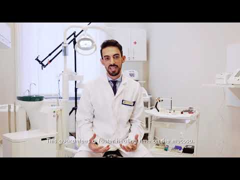
Accelerating Dental Implant Surgery Recovery Time Through Flapless Techniques
The severe reduction in localized surgical trauma correlates directly with an exponentially easier, faster postoperative patient experience. Avoiding scalpel cuts and medical stitches guarantees a significantly faster healing time on the delicate mucosal layer [01:03]. Patients frequently report feeling completely normal and returning to work within just 48 hours of the digital procedure.
Because the actual surgical wound site is essentially a tight, perfectly sealed circle wrapped closely around the titanium healing abutment, the risk of bacterial infiltration drops dramatically. Traditional sutured tissue flaps routinely trap stray food particles and bacterial plaque, leading to painful localized infections. The modern flapless technique inherently provides a sterile, easily maintainable surgical site.
Expected Postoperative Healing Timeline
Dental implant surgery recovery time and tips generally emphasize controlling inflammation, but digital flapless surgery inherently produces very little clinical swelling to begin with. The standard expected healing trajectory for a computer-guided flapless procedure typically looks exactly like this:
- Days 1 to 2: Minor localized tenderness at the circular tissue punch site, easily managed with basic over-the-counter anti-inflammatory medication. No severe external facial bruising or systemic swelling occurs.
- Days 3 to 5: The soft mucosal tissue seals completely around the titanium healing abutment. Patients confidently return to normal daily brushing routines using a soft-bristled toothbrush.
- Weeks 2 to 4: The external mucosal layer is visibly fully healed. Deep internal osseointegration is well underway within the dense cortical jawbone structure.
Choosing the Best Dental Implant Specialist for Complex Oral Surgery
The highly sophisticated nature of digital dentistry still strongly requires a human operator with an extensive clinical background. Specialists who have dedicated uninterrupted years to oral surgery and implantology possess the deep anatomical knowledge required to interpret virtual planning software correctly [00:03]. A computer algorithm simply cannot replace the nuanced clinical judgment needed to handle unexpected physiological variations in bone density.
Locating the absolute best dental implant specialist for complex oral surgery involves rigorously evaluating their documented commitment to modern methodologies. Elite global practitioners actively seek to consistently learn new surgical techniques and continually improve their deep diagnostic knowledge base [00:12]. This academic dedication ensures that traveling patients are treated strictly with the most current, evidence-based surgical protocols available in the industry today.
When actively pursuing international dental tourism, verifying the advanced technological infrastructure of the destination clinic remains paramount. High-end surgical facilities seamlessly integrate CBCT scanners, intraoral optical scanners, and specialized 3D printers into a cohesive digital workflow. Hungary specifically maintains its long-standing reputation as a premier destination because European medical standards strictly ensure that local surgical teams operate efficiently at the highest global tier.
Cost-Effectiveness of Affordable Full Mouth Dental Implants in Europe
Many prospective patients initially assume that computer-guided digital protocols are financially prohibitive due strictly to the expensive imaging technology involved. However, the exact opposite proves true when accurately factoring in long-term clinical stability. The intense physical precision of the customized surgical guide drastically reduces the likelihood of catastrophic implant failure, subsequently preventing the need for incredibly costly secondary revision surgeries.
Securing affordable full mouth dental implants in Europe combines this high-tech digital approach with highly favorable regional economic factors. Clinics operating in central countries like Hungary routinely offer fully guided procedures at a significant fraction of the retail cost seen throughout North America. These substantial financial savings absolutely do not come at the expense of material quality, as the exact same premium global titanium brands and software systems are utilized.
The inherent clinical efficiency of flapless surgery directly lowers the overall final cost by aggressively reducing the total amount of time spent occupying the surgical chair. Shorter clinical appointments automatically translate to lower operational overhead for the clinic, generating direct financial savings that are passed directly to the patient. Investing in a digital guided procedure strictly guarantees that your extensive restorative work is permanently built upon a mathematically perfect titanium foundation.
Ready to Restore Your Smile with Precision?
Discover the benefits of computer-guided dental implants and find out how much you can save on your full mouth restoration by traveling to top-tier clinics abroad.
Get Free QuoteView Full Video Transcript
00:00 [Introductory music and clinic logo visual]
00:03 My name is Dr. Nouri Amir Pasha who has been working at Dentium Implant Center for 2.5 years in the field of oral surgery and implantology.
00:12 In my work i always like to learn new techniques and improve my knowledge.
00:18 What I want to talk about today is a guided implantation which precludes inaccuracy in medical freehand implantation.
00:33 In each case we start with a planning process and we make a condition survey.
00:41 The location and the size of the implants are determined by ct scan, panoramic x-ray and study sample.
00:49 We create a drilling template by a 3D printer.
00:56 With this technique, procedures can be performed without cutting and lobe formation.
01:03 This guarantees a faster healing time on the mucosa.

.png)

.png)

.png)


.png)


Share this listing