


Comprehensive Guide to Surgery Options for a Herniated Disc: Restoring Your Mobility and Relieving Pain
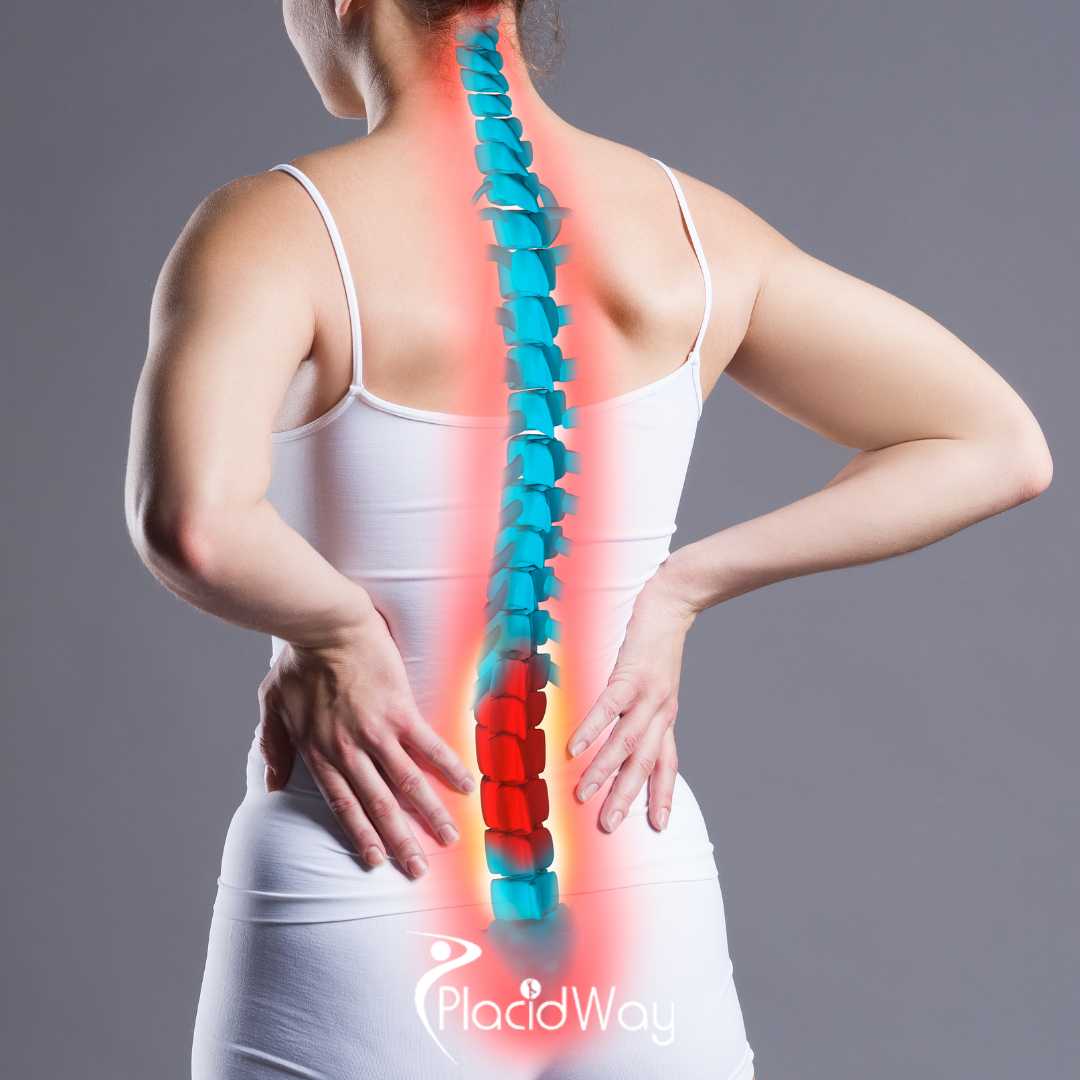
Living with severe, radiating nerve pain can completely derail your quality of life and limit your daily independence. When conservative treatments like targeted physical therapy and epidural steroid injections fail to provide lasting relief, exploring surgery options for a herniated disc becomes a critical next step for many patients. A ruptured or slipped disc can heavily compress delicate spinal nerves, leading to debilitating sciatica, localized numbness, and progressive muscle weakness.
Fortunately, modern medical technology offers highly advanced, minimally invasive disc hernia surgery techniques specifically designed to alleviate this excruciating pain and restore your overall spinal mobility quickly. In this comprehensive medical guide, we will deeply explore the most effective and innovative surgical treatments available in modern orthopedics and neurosurgery today, helping you make an informed decision to reclaim your active, pain-free lifestyle.
Video Chapters & Quick Navigation
Understanding the Anatomy and Pathophysiology of a Herniated Disc
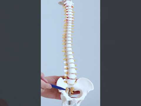
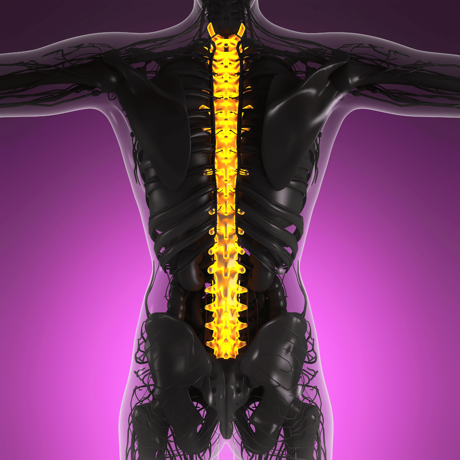
Before diving into the complex surgical solutions, it is absolutely essential to understand the underlying biomechanics of the human spine. The spinal column is composed of individual bones called vertebrae, which are meticulously stacked upon one another. Between each of these bony structures lies an intervertebral disc, which acts as a highly specialized biological shock absorber. These discs allow the spine to bend, twist, and carry the heavy mechanical load of the human body.
The Structure of a Healthy Spinal Disc
A healthy intervertebral disc consists of two primary anatomical components. The outer ring, known medically as the annulus fibrosus, is a tough, durable, and fibrous exterior layer designed to protect the inner core. The inner core, called the nucleus pulposus, is a soft, gel-like substance that provides the vital hydration and cushioning needed for impact absorption. As we age, these discs naturally undergo degenerative changes, losing their crucial hydration and becoming increasingly susceptible to structural tears and painful ruptures.
How a Herniation Occurs
A herniated disc occurs when the tough outer layer (annulus fibrosus) sustains a physical tear, allowing the soft, jelly-like inner core (nucleus pulposus) to extrude outwards into the spinal canal. As highlighted in the video at , the spinal canal has a very limited amount of free space. When disc material invades this tightly enclosed area, it directly compresses the highly sensitive spinal cord or the exiting nerve roots. This intense mechanical compression, combined with the severe chemical inflammation caused by the extruded gel, creates the agonizing pain profile commonly associated with a slipped disc.
Recognizing When Surgery Options for a Herniated Disc Become Necessary
The vast majority of patients diagnosed with a herniated disc will not require immediate surgical intervention. Medical guidelines heavily suggest that patients first undergo an exhaustive, comprehensive trial of conservative therapies. This non-surgical approach typically includes targeted physical therapy to strengthen core musculature, non-steroidal anti-inflammatory drugs (NSAIDs), targeted epidural steroid injections to reduce nerve swelling, and modified activity levels.
The Failure of Conservative Treatment
Surgeons generally recommend exploring slipped disc treatment options only after a patient has actively participated in conservative care for a period of six to twelve weeks without experiencing significant symptomatic relief. If the debilitating pain continues to negatively interfere with basic daily activities, such as walking, sitting, or sleeping, the mechanical compression is unlikely to resolve on its own. At this juncture, continuing to delay surgical intervention can lead to long-term chronic pain syndromes and severe nerve degradation.
Medical Red Flags Requiring Immediate Surgery
While patience is a virtue in spinal recovery, certain neurological "red flag" symptoms mandate urgent, if not emergency, surgical evaluation. As noted in the discussion at , sudden and severe muscle weakness, such as foot drop, requires immediate medical attention. More critically, the sudden onset of bowel or bladder incontinence, accompanied by saddle anesthesia (numbness in the groin area), indicates Cauda Equina Syndrome. This is an absolute medical emergency where emergency decompressive surgery is required to prevent permanent, irreversible paralysis.
The Gold Standard: Lumbar Microdiscectomy Procedure
For patients suffering from severe, unrelenting sciatica caused by a herniated disc in the lower back, the lumbar microdiscectomy procedure has long been established as the absolute gold standard in modern spine surgery. This highly refined operation is specifically engineered to achieve maximum nerve decompression while simultaneously minimizing damage to the surrounding healthy spinal tissues and musculature.
The Surgical Technique
During a microdiscectomy, the board-certified neurosurgeon or orthopedic spine specialist creates a very small incision, typically less than an inch long, directly over the affected spinal level. Utilizing specialized tubular retractors, the surgeon gently pushes the back muscles aside rather than cutting violently through them. A high-powered surgical microscope is then brought into the sterile field, providing unparalleled, brightly illuminated magnification of the delicate neural structures.
Once the compressed nerve root is carefully identified and gently retracted to safety, the surgeon utilizes micro-instruments to meticulously grasp and extract only the herniated portion of the disc. The healthy, unruptured portion of the disc is left completely intact to maintain proper spinal biomechanics and height. This precision-driven approach boasts an exceptional success rate, frequently reported at over 90% for complete sciatica pain relief surgery.
| Surgical Feature | Traditional Open Discectomy | Minimally Invasive Microdiscectomy |
|---|---|---|
| Incision Size | Large (2 to 4 inches) | Very Small (less than 1 inch) |
| Muscle Disruption | Extensive cutting and stripping | Gentle dilation and tissue sparing |
| Hospital Stay | 1 to 3 days inpatient | Often outpatient (same-day discharge) |
| Recovery Speed | Slower, more post-operative pain | Rapid, highly accelerated recovery |
Endoscopic Spine Surgery for Herniated Disc: The Ultra-Minimally Invasive Approach
While microdiscectomy is highly effective, the rapid evolution of medical technology has birthed an even less invasive alternative. Endoscopic spine surgery for a herniated disc represents the absolute bleeding edge of modern neurosurgical innovation. This state-of-the-art procedure is fundamentally transforming how spine specialists approach lumbar disc herniations, offering a pathway to pain relief with almost zero collateral tissue damage.
How Endoscopic Discectomy Works
Instead of relying on a traditional surgical microscope and open retractors, the endoscopic approach utilizes a high-definition, fiber-optic camera known as an endoscope. The surgeon creates an incredibly tiny incision, often no larger than an eight-millimeter keyhole puncture. As detailed at , the endoscope is seamlessly navigated through the natural anatomical openings in the spine, bypassing complex muscle layers entirely.
The surgeon watches a large, high-resolution monitor in real-time as they operate. Utilizing microscopic grasping tools, precision radiofrequency probes, and occasionally targeted holmium lasers routed directly through the hollow center of the endoscope, the surgeon pinpoints and vaporizes the exact piece of herniated disc material. A continuous flow of saline water runs through the endoscope to keep the surgical field pristinely clear and actively cool the surrounding nerve tissues.
The Benefits of "Awake" Spine Surgery
One of the most profound advantages of endoscopic spine surgery is the anesthesia protocol. Many leading spine centers now perform these advanced procedures under local anesthesia with light twilight sedation, rather than risky general anesthesia. This means the patient is awake, comfortable, and able to provide real-time neurological feedback to the surgeon during the operation. This hyper-advanced technique practically eliminates the risk of anesthesia-related complications and allows patients to walk out of the surgical center merely hours after the procedure concludes.
Laminectomy and Laminotomy: Decompressing the Spinal Canal
In certain complex clinical scenarios, a herniated disc is not the sole culprit causing the patient's severe radiating pain. As the human spine naturally ages, patients frequently develop concurrent conditions such as severe spinal stenosis, hypertrophic ligament thickening, or aggressive osteophyte (bone spur) formations. When a massive disc herniation is combined with these degenerative factors, simply removing the soft disc material is entirely insufficient to fully free the heavily compressed nerves.
Creating Vital Anatomical Space
To adequately decompress the spinal canal, a surgeon may elect to perform a laminectomy or a more conservative laminotomy alongside the discectomy. The lamina is the bony, roof-like arch located at the posterior (back) of the vertebra that covers and protects the delicate spinal canal. During a full laminectomy, the surgeon completely removes this bony arch to drastically widen the internal diameter of the spinal canal.
Conversely, a laminotomy is a slightly more conservative, tissue-sparing approach. Instead of removing the entire bony structure, the surgeon utilizes a high-speed surgical burr to meticulously shave away a small, strategic window in the lamina. This targeted bone removal creates just enough microscopic space to safely extract the hidden disc herniation and drastically relieve the suffocating pressure placed directly on the exiting nerve root. Both techniques are highly effective in treating complex, multi-factorial radiculopathy.
Artificial Disc Replacement Surgery: Preserving Natural Spinal Motion
Historically, when a spinal disc was completely destroyed by a massive herniation or severe degenerative disc disease, the only viable, long-term surgical solution was a traditional spinal fusion. While fusion successfully eliminated the excruciating pain, it came at the heavy cost of permanently locking the affected vertebrae together, thereby eliminating all natural motion at that specific spinal segment. Today, artificial disc replacement surgery presents a revolutionary, motion-preserving alternative.
The Biomechanics of an Artificial Disc
Also known as total disc arthroplasty, artificial disc replacement surgery involves the complete removal of the diseased and herniated disc. Once the damaged disc space is thoroughly cleared and the spinal nerves are completely freed from compression, the surgeon precisely implants a high-tech prosthetic device. Mentioned in the video at , these advanced implants are engineered from durable medical-grade materials, such as heavy-duty cobalt-chromium alloys and highly durable ultra-high-molecular-weight polyethylene.
The intricate design of the artificial disc intimately mimics the six degrees of freedom found in a healthy, biological human disc. It seamlessly allows for flexion, extension, lateral bending, and natural axial rotation. By successfully maintaining the natural biomechanics of the spine, this innovative procedure drastically reduces the mechanical stress placed on the adjacent, neighboring spinal levels.
Ideal Candidates for Disc Arthroplasty
It is important to aggressively note that not every patient is a suitable candidate for artificial disc replacement. Ideal candidates are generally younger to middle-aged adults suffering from symptomatic single-level disc herniations who possess excellent, healthy bone density. Patients suffering from significant facet joint arthritis, severe osteoporosis, or pre-existing spinal instability are generally disqualified from this procedure and must pursue alternative surgical routes to ensure long-term clinical safety.
Spinal Fusion for Ruptured Discs: Achieving Long-Term Spinal Stability
Despite the rapid rise of motion-preserving technologies, traditional spinal fusion for a ruptured disc remains an incredibly vital and frequently necessary surgical intervention. When a severe disc herniation is accompanied by severe mechanical instability, such as spondylolisthesis (where one vertebra forcefully slips over another), simply removing the disc material would leave the spine dangerously unstable. In these complex cases, fusion is absolutely required to permanently halt painful micro-movements.
The Mechanics of Spinal Fusion
During a comprehensive spinal fusion procedure, the surgeon meticulously removes the entirety of the degenerated disc to thoroughly decompress the affected nerve roots. A highly specialized spacer, often packed densely with biologically active bone graft material, is firmly inserted into the newly empty disc space. This essential bone graft can be harvested directly from the patient's own pelvis (autograft) or safely obtained from a sterile donor bone bank (allograft).
To ensure absolute mechanical rigidity while the slow biological fusion process takes place, the surgeon heavily reinforces the spine using medical-grade titanium pedicle screws and connecting stabilizing rods. Over the course of several months, the living bone graft triggers massive cellular bone growth, permanently welding the two separate vertebrae into one solid, continuous, and incredibly stable bone mass.
Different Approaches to Fusion
Modern spine surgeons can approach the spine from multiple intricate angles to perform a highly successful fusion, tailoring the operation to the patient's exact anatomical needs. These surgical approaches include:
- ALIF (Anterior Lumbar Interbody Fusion): The surgeon approaches the spine through the abdomen, completely avoiding traumatic disruption of the posterior back muscles.
- PLIF (Posterior Lumbar Interbody Fusion): The traditional approach, accessing the spine directly from the back, allowing for excellent visualization of the nerve roots.
- TLIF (Transforaminal Lumbar Interbody Fusion): A lateralized approach from the back that requires less nerve retraction, drastically reducing the risk of post-operative nerve damage.
- LLIF (Lateral Lumbar Interbody Fusion): Approaching the spine directly through the patient's side, utilizing advanced minimally invasive tubular retractors.
Navigating the Comprehensive Recovery Time for Herniated Disc Surgery
Understanding the realistic recovery time for herniated disc surgery is essential for managing patient expectations and ensuring a successful, complication-free outcome. Recovery timelines vary drastically depending on the specific surgical approach utilized, the patient's pre-operative physical health, and their absolute dedication to strict post-operative rehabilitation protocols.
The Immediate Post-Operative Phase (Weeks 1-2)
For patients undergoing a minimally invasive lumbar microdiscectomy or an advanced endoscopic procedure, the initial recovery is remarkably swift. As noted at , most patients report immediate, profound relief from their agonizing sciatica pain upon waking up in the recovery room. During the first two weeks, patients are heavily encouraged to engage in frequent, short, and gentle walks to continuously promote healthy blood circulation and aggressively prevent deep vein thrombosis (blood clots).
However, patients must strictly adhere to the universal "BLT" restrictions: Absolutely No Bending, No Lifting (anything heavier than a gallon of milk), and No Twisting of the spine. Ignoring these critical bio-mechanical restrictions can easily result in a catastrophic re-herniation, requiring an immediate secondary operation.
Physical Therapy and Long-Term Healing (Weeks 3-12)
Around the third or fourth post-operative week, the vast majority of surgeons will officially clear the patient to begin a highly structured, medically supervised physical therapy regimen. The primary, critical goal of this therapy is to aggressively strengthen the deep core musculature and the intricate paraspinal muscles that support the delicate spinal column. By developing a strong, muscular "internal corset," patients heavily reduce the long-term load placed directly on the vulnerable intervertebral discs.
While patients with physically demanding, labor-intensive jobs may require up to three full months of continuous healing before safely returning to full duty, desk workers can often resume their normal professional activities within two to three weeks, provided they utilize highly ergonomic seating and take frequent standing breaks.
Global Healthcare Solutions and Medical Tourism for Spine Surgery
The financial realities of complex, high-tech orthopedic spine surgery can be incredibly daunting. In many Western nations, specifically the United States, out-of-pocket costs for advanced procedures like artificial disc replacement surgery or multi-level spinal fusion can easily exceed fifty thousand dollars, even for patients possessing comprehensive health insurance policies. This exorbitant financial burden has fueled a massive, global rise in medical tourism for spine-related conditions.
Accessing World-Class Care Internationally
Patients are increasingly looking beyond their domestic borders to secure highly affordable, world-class medical care. Nations such as Mexico, Turkey, Germany, and South Korea have invested billions of dollars in establishing ultra-modern, internationally accredited surgical hospitals. These premium medical facilities are frequently staffed by highly decorated, internationally trained neurosurgeons who hold prestigious board certifications from top-tier medical institutions in the US and the UK.
Through dedicated medical facilitation networks like PlacidWay, patients suffering from severe spinal conditions can easily access heavily discounted, comprehensive surgical packages. These all-inclusive, premium packages routinely cover the cost of the advanced surgical procedure, state-of-the-art titanium implants, lengthy hospital stays, pre-operative MRI imaging, and luxurious hotel accommodations for the duration of the critical recovery period, often resulting in total savings of up to seventy percent compared to domestic medical billing rates.
Ready to Explore Your Spine Surgery Options?
Do not let debilitating nerve pain permanently dictate the terms of your life. PlacidWay Medical Tourism seamlessly connects you with elite, internationally recognized spine surgeons offering world-class disc hernia surgery at highly affordable prices. Let our experts guide your journey back to a pain-free life.
GET A FREE MEDICAL QUOTE TODAY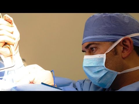




.png)









Share this listing