Comprehensive Guide to Ganglion Cyst Treatment and Surgical Removal
Noticing a sudden, unexplained lump on your wrist or hand can be a source of immediate concern. However, in the vast majority of cases, these localized swellings are diagnosed as benign fluid-filled sacs that develop along joints or tendons. If you are seeking effective ganglion cyst treatment, understanding what these cysts are and how they function is your first vital step. This comprehensive medical guide explores the exact nature of these common non-cancerous lumps, identifying why they form, the warning signs to watch for, and the complete spectrum of advanced medical treatments available today.
Video Chapters & Quick Navigation
What Exactly is a Ganglion Cyst?
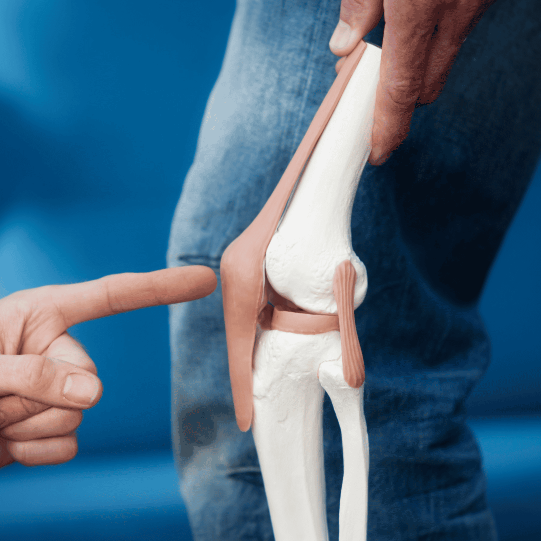
A ganglion cyst is an incredibly common, benign tumor or swelling that typically forms on top of a joint or the covering of a tendon. To visualize this, imagine a tiny, fluid-filled balloon attached to a hollow stalk that connects directly to the underlying tissues. The word "ganglion" translates to knot, which accurately describes the knot-like mass that develops beneath the surface of the skin. These cysts are fundamentally non-cancerous and will never spread to other areas of the body or transform into malignant tumors.
Inside this sac-like structure is a thick, sticky, clear, and colorless jelly-like material known as synovial fluid. Your body naturally produces synovial fluid to lubricate and nourish your joints and tendon sheaths, allowing for smooth, frictionless movement. However, when a weakness occurs in the joint capsule or tendon sheath, this fluid can leak out and accumulate in a concentrated sac, resulting in the visible lump associated with ganglion cysts.
Depending on their size and the exact location, these cysts may feel incredibly firm to the touch, or they may feel somewhat spongy and mobile. It is important to note that the size of a ganglion cyst is rarely static. Because the cyst is connected to the joint space by a one-way valve or stalk, joint movement can literally pump more fluid into the cyst, causing it to swell and increase in size during periods of heavy physical activity or repetitive motion.
Common Anatomical Locations for Cyst Formation
While a ganglion cyst can technically develop near any joint in the human body, clinical data shows they have a strong predilection for specific anatomical regions, primarily due to the complex biomechanics and high stress load placed on these particular joints.
The Dorsal Wrist (Back of the Wrist)
The vast majority of ganglion cysts—approximately 60 to 70 percent—develop on the dorsal aspect, or the back of the wrist. They typically originate from the scapholunate joint, which is a critical connection between two small carpal bones in the wrist. When a patient bends their wrist forward, these dorsal cysts become much more prominent and visually noticeable.
The Volar Wrist (Palm Side of the Wrist)
The second most common location is the volar wrist, occurring on the palm side, often near the base of the thumb or where you would normally feel your radial pulse. Cysts in this area require careful evaluation by an orthopedic specialist, as they intimately wrap around or press against the radial artery and various vital nerves, making both non-surgical and surgical treatments slightly more complex.
The Base and Tips of the Fingers
Ganglion cysts can also formulate at the base of the fingers on the palm side. These are often referred to as flexor tendon sheath cysts and typically present as small, hard bumps about the size of a pea. When they occur at the fingertip just below the cuticle, they are called mucous cysts. Mucous cysts are heavily associated with osteoarthritis in the distal interphalangeal (DIP) joint and can occasionally cause a visible depression or groove in the growing fingernail.
Underlying Causes and Hidden Risk Factors
Despite extensive medical research into orthopedic pathology, the exact underlying mechanism that triggers the initial formation of a ganglion cyst remains somewhat elusive. However, the prevailing medical theory is known as the "micro-trauma" hypothesis. This theory suggests that repeated stress, friction, or minor unnoticed injuries to a joint capsule or tendon sheath cause the connective tissue to break down or weaken locally.
As this tissue weakens, the synovial fluid that normally bathes the joint leaks through the micro-tears. Because the surrounding tissue attempts to contain this rogue fluid, it forms a localized sac or cyst wall. Over time, the one-way valve effect draws more fluid out of the joint and traps it in the cyst.
While anyone can develop these localized swellings, several distinct risk factors heavily influence their occurrence. Understanding these risk factors is an important element in the accurate diagnosis of wrist lumps:
- Age and Gender Demographics: Ganglion cysts are overwhelmingly more common in women than in men, occurring at a ratio of nearly 3 to 1. Furthermore, they are most frequently diagnosed in patients between the ages of 20 and 40 years old.
- Joint and Tendon Injuries: Patients who have suffered previous trauma to a joint, such as a severe sprain, fracture, or hyperextension injury, are significantly more likely to develop cysts in that exact location in the future.
- Occupational and Athletic Stress: Repetitive joint stress is a massive contributing factor. Gymnasts, tennis players, weightlifters, and individuals who type extensively are prone to these cysts due to the constant mechanical load placed on the wrist joints.
- Osteoarthritis: In older demographics, wear-and-tear arthritis in the finger joints nearest the fingernails is the primary driver for the development of mucous cysts.
Recognizing the Signs and Symptoms of Wrist Cysts
The clinical presentation of a ganglion cyst can vary dramatically from one patient to the next. For many individuals, the only symptom is a visible, cosmetic deformity—a painless lump that causes aesthetic concern but no functional impairment. The lump typically appears suddenly, though it can also develop gradually over several months.
The size of the mass is a primary characteristic. Most cysts are roughly the size of a small pea, while others can grow to the size of a golf ball. It is highly characteristic for a ganglion cyst to fluctuate in size. Patients frequently report that the lump gets larger and tighter after heavy activity, and shrinks after periods of prolonged rest.
However, when a cyst places mechanical pressure on adjacent anatomical structures, it ceases to be asymptomatic. If the cyst grows near a nerve, patients may experience localized or radiating pain, tingling sensations, numbness, or a deep aching throb that worsens with joint movement. If the cyst compresses a tendon, the patient may experience noticeable muscle weakness or restricted range of motion, making simple tasks like gripping a steering wheel or opening a jar exceedingly difficult and painful.
Advanced Diagnostic Procedures for Ganglion Cysts
Accurate diagnosis is paramount before embarking on any ganglion cyst treatment plan. While the visual appearance is often a dead giveaway for an experienced orthopedic hand specialist, medical professionals must rule out other potential causes of wrist lumps, such as lipomas, giant cell tumors of the tendon sheath, or vascular aneurysms.
The diagnostic process typically begins with a thorough medical history and a physical examination. The physician will apply gentle pressure to the mass to test for tenderness and assess its consistency. Because the cyst is filled with translucent fluid, a classic diagnostic technique called transillumination is frequently employed. By shining a penlight directly against the mass in a darkened room, the physician can observe if the light passes through it. If the mass glows, it confirms the presence of clear fluid, heavily pointing to a ganglion cyst.
| Diagnostic Tool | Primary Purpose | When It Is Used |
|---|---|---|
| Physical Exam & Transillumination | To check mass consistency and confirm the presence of clear fluid. | Standard for almost all initial consultations and visible lumps. |
| Diagnostic Ultrasound | To visualize fluid-filled sacs and evaluate blood flow (vascularity). | To differentiate cysts from solid tumors or to guide needle aspirations. |
| Magnetic Resonance Imaging (MRI) | To provide detailed, cross-sectional 3D images of soft tissues. | Used for "occult" (hidden) cysts causing unexplained wrist pain. |
For cysts that are too small to see but are causing severe pain—known as occult ganglion cysts—advanced imaging becomes necessary. Ultrasound is a quick, inexpensive, and highly effective way to differentiate a fluid-filled cyst from a solid soft-tissue tumor. If the diagnosis remains unclear, an MRI serves as the gold standard for soft tissue evaluation, offering unparalleled detail of the cyst's exact size, location, and its relationship to surrounding nerves and ligaments.
Effective Non-Surgical Treatment for Ganglion Cysts
The approach to non-surgical treatment for ganglion cysts is highly dependent on the patient's symptoms. If the lump is completely painless and does not interfere with daily activities, the most common medical recommendation is simple watchful waiting. Medical studies indicate that up to 50 percent of ganglion cysts may spontaneously resolve and disappear on their own without any medical intervention, as the fluid is naturally reabsorbed by the body.
Immobilization and Splinting
Because physical activity and joint movement actively pump more synovial fluid into the cyst, temporary immobilization is a highly logical first-line treatment. By prescribing a custom wrist brace or splint, the physician aims to restrict movement, shut off the fluid pump mechanism, and allow the cyst to shrink. As the mass decreases in size, pressure is relieved from surrounding nerves, rapidly alleviating pain and discomfort.
The Aspiration Procedure
If immobilization fails or if the cyst is causing severe aesthetic or physical distress, a physician may recommend a clinical procedure known as aspiration. This is an outpatient procedure performed under local anesthesia. The doctor sterilizes the area, injects a numbing agent, and then inserts a hollow needle directly into the cyst.
Once the needle breaches the sac, the thick, gelatinous fluid is drawn out into a syringe, causing the visible lump to collapse immediately. To reduce local inflammation and help seal the cyst wall, the physician may inject a corticosteroid medication into the empty sac before removing the needle. While aspiration is quick, painless, and provides instant relief, patients must be aware of the recurrence rates. Because aspiration only drains the fluid but leaves the root "stalk" and the cyst lining intact, there is an estimated 50 to 70 percent chance that the cyst will refill with fluid over time.
Ganglion Cyst Removal Surgery: When Excision is Necessary
When conservative measures fail, when a cyst repeatedly returns after multiple aspirations, or when the mass is causing severe nerve compression and functional loss, surgical excision becomes the definitive treatment. Ganglion cyst removal surgery is a precise orthopedic procedure designed not just to drain the fluid, but to completely remove the anatomical structures causing the problem.
The goal of surgery is to meticulously separate the cyst from the surrounding blood vessels, nerves, and tendons. More importantly, the surgeon must trace the cyst down to its base and remove the stalk, along with a small portion of the involved joint capsule or tendon sheath. By removing the root cause, the recurrence rate drops dramatically to approximately 5 to 15 percent.
Open Excision vs. Arthroscopic Surgery
Depending on the cyst's location and the surgeon's expertise, the procedure can be performed using traditional open surgery or minimally invasive arthroscopic techniques.
- Open Surgery: The surgeon makes an incision directly over the cyst. This provides direct visualization, allowing the surgeon to carefully dissect the mass away from the radial artery or median nerve. It is highly effective but results in a slightly larger scar and potentially a longer recovery period for the surrounding tissues.
- Arthroscopic Surgery: Often used for dorsal wrist cysts. The surgeon makes two tiny incisions (portals). A microscopic camera is inserted into one, and specialized miniature cutting tools into the other. The cyst stalk is removed from the inside of the joint. This approach preserves the joint capsule's outer layers, minimizes scarring, and often accelerates post-operative rehabilitation.
Post-Operative Recovery and Rehabilitation
Ganglion cyst removal is typically performed as an outpatient procedure, meaning you will return home the very same day. Following the surgery, your hand and wrist will be bandaged with a bulky dressing and frequently supported by a splint to immobilize the joint for the first week. This initial immobilization is crucial to allow the surgical incision and the internal joint capsule to heal properly without being stretched by movement.
Pain is generally mild to moderate and can be managed effectively with over-the-counter analgesics and localized ice therapy. Patients are encouraged to keep their hand elevated above heart level for the first 48 hours to minimize throbbing and post-operative swelling. Suture removal usually occurs 10 to 14 days after the operation.
Once the splint is removed, the rehabilitation phase begins. A structured physical therapy program is highly recommended. Physical therapists will guide patients through active and passive range-of-motion exercises to prevent joint stiffness and scar tissue formation. Furthermore, scar massage techniques are taught to ensure the healing tissue remains pliable. Most patients can return to normal daily activities within two to four weeks, though heavy lifting and rigorous sports should be avoided for up to six weeks to guarantee complete internal healing.
Navigating the Cost of Orthopedic Care
While the clinical path to treating a ganglion cyst is straightforward, the financial realities of orthopedic care can be a significant hurdle for many patients. In the United States, the United Kingdom, and parts of Europe, the cost of specialized hand surgery, pre-operative MRIs, facility fees, anesthesia, and post-operative physical therapy can accumulate into a massive financial burden, especially for patients with high deductibles or those lacking comprehensive health insurance.
This financial barrier has driven a massive surge in medical tourism for orthopedic procedures. Patients are increasingly looking globally for high-quality, specialized care that does not compromise on safety or medical expertise but offers a fraction of the domestic price tag. Reputable international medical centers boast board-certified orthopedic surgeons, state-of-the-art diagnostic imaging, and dedicated rehabilitation wings, providing end-to-end care that rivals the best domestic hospitals.
Seeking international orthopedic solutions allows patients to regain their joint function and live pain-free without devastating their finances. With dedicated medical tourism coordinators managing everything from surgical consultations to post-operative hotel stays, the journey to wellness is streamlined, accessible, and deeply focused on patient recovery and satisfaction.
Explore Affordable Global Orthopedic Solutions
Don't let the high cost of medical care keep you living in pain or discomfort. PlacidWay connects you with globally accredited orthopedic clinics offering world-class ganglion cyst treatments and excision surgeries at a fraction of the cost. Let us help you find the right specialist and manage your entire medical journey.
REQUEST A FREE CONSULTATION
.png)


Share this listing