Finding specialized care for diabetes-induced vision issues is highly critical, and seeking ocular paralysis treatment in Mexico has become a top viable option for international patients. Ocular paralysis, particularly in individuals struggling with severe diabetes, is a complex neurological and ophthalmological condition that significantly impairs daily life. High systemic blood sugar levels can silently and progressively damage the delicate cranial nerves responsible for precise eye movement, leading to distressing symptoms like binocular double vision and severe muscle weakness.
Understanding the intricate mechanisms behind diabetic ocular paralysis, often referred to medically as diabetic cranial nerve palsy, allows patients to make highly informed decisions regarding their personal healthcare. Exploring the specific diagnostic procedures, the profound importance of strict blood glucose regulation, and the advanced therapeutic options available equips individuals with the necessary knowledge to effectively manage their condition. Knowing exactly how medical professionals approach this delicate diagnosis empowers the journey toward fully restored vision and long-term neurological health.
Video Chapters
- What is Diabetic Ocular Paralysis?
- The Link Between Blood Sugar and Nerve Damage
- Recognizing Symptoms: Double Vision
- Diagnosing Cranial Nerve Palsy in Diabetics
- Excluding Other Neurological Conditions
- Effective Ocular Paralysis Treatment Protocols
- Nerve Regeneration Therapies and Recovery
- Long-Term Prevention for Eye Complications
- Understanding Financial Benefits Abroad
- Why Choose Care in Specialized Clinics
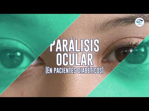
What Exactly is Diabetic Ocular Paralysis?
Dr. Juan Pablo Rodríguez, an experienced ophthalmic surgeon, clearly defines this condition as a partial or complete loss of control of the extraocular muscles. According to his professional explanation in the video, this functional deficit fundamentally alters how the eye operates within its socket [00:16]. When a patient develops diabetic ocular paralysis, the intricate coordination between the two eyes is massively disrupted. The extraocular muscles are uniquely responsible for precise eye movements, allowing individuals to successfully track objects and maintain a steady gaze without physical strain.
These specific muscles rely heavily on constant electrical signals originating from the brain, meticulously transmitted through specific cranial nerves. A sudden disruption in any of these neurological pathways leads to immediate visual disturbances that strongly require prompt medical evaluation. For diabetic patients, the overall risk of experiencing this exact type of neurological interruption is significantly higher due to systemic metabolic issues that aggressively degrade vascular health over an extended period.
The acute loss of muscle control is very rarely an isolated clinical problem; it serves as a critical indicator of broader systemic distress occurring within the body. When the eye muscles fail to receive appropriate electrical signals, the affected eye may drift inward, outward, upward, or downward entirely unexpectedly. This specific ocular misalignment is not simply a cosmetic issue but a profound, debilitating functional impairment. Daily activities such as reading, driving, and basic walking can become highly hazardous due to heavily compromised depth perception and completely altered spatial awareness.
The Underlying Link Between High Blood Sugar and Nerve Damage
The fundamental physical trigger for cranial nerve palsy in diabetic patients heavily lies in poor metabolic management over extended periods. The condition is directly caused by a completely inadequate control of glucose or sugar levels circulating within the body [00:25]. Chronic hyperglycemia inflicts severe, often irreversible damage on the delicate vascular network that constantly supplies vital blood to peripheral and cranial nerves. This microscopic network, medically known as the vasa nervorum, is extremely vulnerable to toxic, daily fluctuations in blood sugar.
When blood sugar levels remain highly elevated, these vital small blood vessels undergo detrimental, permanent structural changes. They become excessively stiff, dangerously narrowed, and significantly less capable of safely delivering essential oxygen and nutrients to the surrounding nerve tissues. This pathological state, known precisely as diabetic microvascular ischemia, essentially starves the fragile nerve fibers. Without adequate and continuous blood flow, the specific nerve responsible for actively stimulating the normal functioning of the eye muscles becomes heavily compromised [00:30].
Additionally, persistently high glucose levels trigger dangerous, toxic biochemical pathways deeply within the neurological system. The abnormal, rapid accumulation of sorbitol within nerve cells aggressively draws in excess water, leading to severe cellular swelling and massive structural degradation. Unmanaged oxidative stress simultaneously generates harmful free radicals that aggressively attack the myelin sheath, the protective biological coating of the nerves. This intense demyelination process severely slows down the vital transmission of electrical impulses, eventually culminating in full, debilitating muscle paralysis.
Recognizing the Primary Symptoms: Double Vision and Muscle Weakness
One of the absolutely most disorienting and immediate symptoms of ocular paralysis is the sudden, jarring onset of double vision. Because the affected eye cannot physically align properly with the healthy eye, the brain abruptly receives two distinctly different, completely conflicting visual signals [00:21]. This phenomenon, widely recognized as diplopia, is extremely distressing and highly disorienting for affected patients. The human brain struggles immensely to artificially fuse these mismatched images, resulting in a confusing, nauseating visual experience that disrupts all normal daily activities.
Actively seeking effective double vision treatment for diabetes successfully begins with pinpointing the exact nature of the visual disturbance. Binocular diplopia, which strictly occurs only when both eyes are completely open, is the definitive, undeniable hallmark of extraocular muscle paralysis. If the patient physically covers one eye, the double vision instantly and fully disappears, providing a highly critical diagnostic clue for the physician. Depending entirely on which specific cranial nerve has sustained ischemic damage, the double vision may continuously present as horizontal, vertical, or even a diagonal misalignment.
| Cranial Nerve | Primary Ocular Function | Classic Symptoms of Paralysis |
|---|---|---|
| Cranial Nerve III (Oculomotor) | Controls most eye movements, pupil constriction, and eyelid elevation | Down and out eye deviation; Severe drooping eyelid (ptosis); Diagonal double vision |
| Cranial Nerve IV (Trochlear) | Controls downward and inward eye movement primarily | Upward eye deviation; Worsening double vision when looking down or reading; Constant head tilt to compensate |
| Cranial Nerve VI (Abducens) | Controls outward eye movement (abduction) away from the nose | Inward eye deviation (esotropia); Worsening horizontal double vision when looking toward the affected side |
Beyond the immense frustration of double vision, patients very frequently report a sudden onset of severe, localized aching pain around or directly behind the affected eye. This deeply localized pain very often precedes the actual visible paralysis by several days and is strongly correlated directly with the acute ischemic injury occurring deep within the nerve bundle. In severe cases involving the third cranial nerve, individuals may also heavily experience a dramatic, complete drooping of the upper eyelid, clinically referred to as ptosis, which visually occludes the eye completely.
The Diagnostic Process for Diabetic Cranial Nerve Palsy
Accurate, timely identification of this intricate neurological issue heavily requires a highly comprehensive evaluation by a deeply qualified eye care specialist. The definitive diagnosis of this specific muscular paralysis is typically established directly in the consulting room through a highly detailed ophthalmological exam for eye paralysis [00:37]. The attending physician will always begin by taking an incredibly exhaustive medical history, focusing particularly heavily on the patient's long-term diabetes management, recent hemoglobin A1c readings, and the exact chronological timeline of symptom progression.
During the active physical examination phase, the specialized ophthalmologist meticulously assesses the extraocular movements across absolutely all fundamental fields of gaze. By carefully instructing the patient to visually track a moving target without moving their head, the specialist can easily isolate and confidently identify which specific ocular muscle is heavily underperforming. The standard cover-uncover test and the alternate cover test are actively utilized to carefully measure the exact optical degree of ocular deviation. These highly precise optical measurements are incredibly crucial for establishing a reliable, solid baseline to accurately monitor future diabetic ocular paralysis recovery.
Another absolutely vital, non-negotiable component of the clinical evaluation is the meticulous, bright-light assessment of pupillary function and reactivity. In the vast, overwhelming majority of cases involving diabetic third nerve palsy, the patient's pupil strongly remains entirely normal in size and highly reactive to bright light stimulation. This specific phenomenon, widely known medically as pupil-sparing, actively serves as a remarkably strong clinical indicator that the paralysis directly stems from diabetic microvascular ischemia rather than a significantly more dangerous compressive brain lesion.
Excluding Other Potential Neurological Conditions Safely
One of the genuinely significant clinical advantages of diagnosing diabetic-related cranial nerve palsies is the relatively highly straightforward, non-invasive diagnostic pathway. In the vast majority of these presented cases, the condition strictly does not require additional, highly expensive imaging tests, provided the overall clinical picture strongly and undeniably points toward metabolic diabetes [00:41]. If an individual possesses a solidly known history of poorly controlled diabetes and actively presents with classic pupil-sparing symptoms, the clinical diagnosis is extremely confident based entirely on the physical exam alone.
However, specialized physicians remain exceptionally highly vigilant and extraordinarily cautious regarding any atypical neurological presentations during the exam. The incredibly notable exception to the standard diagnostic rule occurs precisely when the examining physician actively suspects that the acute paralysis is not directly caused by underlying diabetes. If a patient suddenly experiences a severe, thunderclap headache, if multiple cranial nerves simultaneously become heavily involved, or if the pupil suddenly becomes abnormally enlarged and totally unresponsive, immediate neuroimaging protocols immediately become mandatory.
Severe, life-threatening medical conditions such as rapidly expanding intracranial aneurysms, particularly those dangerously situated on the posterior communicating artery, can heavily compress the third cranial nerve and generate perfectly identical paralytic symptoms. Growing brain tumors, acute ischemic strokes, or severe autoimmune inflammatory conditions like myasthenia gravis absolutely must be definitively ruled out in atypical, complex patient cases. Magnetic Resonance Imaging (MRI) or specialized Computed Tomography (CT) scans are deployed extraordinarily rapidly when these dangerous non-diabetic root causes are actively suspected.
Effective Ocular Paralysis Treatment Protocols in Practice
Successfully managing this complex ophthalmic condition strictly requires a dedicated, dual-pronged medical approach, actively addressing both the immediate visual discomfort and the incredibly dangerous underlying metabolic crisis. The absolute primary and undisputedly most effective method to reliably remedy ocular paralysis is by achieving incredibly strict, unwavering daily control over blood glucose levels [00:48]. This intense systemic, whole-body approach is entirely non-negotiable; without fully and permanently stabilizing blood sugar, further severe nerve damage is exceptionally highly likely, and any potential physical recovery will be massively delayed.
Endocrinologists and dedicated primary care physicians must actively work collaboratively and incredibly closely with the affected patient to continuously optimize their daily diabetes management plan. This highly comprehensive strategy very often involves carefully adjusting synthetic insulin dosages, introducing highly effective new oral medications, or strictly enforcing much stricter dietary carbohydrate protocols. Utilizing advanced continuous glucose monitoring devices can comfortably provide highly valuable, continuous real-time metabolic data to successfully prevent the drastic glucose spikes that continually exacerbate active nerve ischemia.
To highly actively manage the absolutely debilitating double vision during the prolonged, frustrating recovery phase, leading ophthalmologists consistently employ conservative, completely non-invasive optical strategies. The absolute simplest, highly effective method is standard occlusion therapy, which carefully involves securely placing an opaque patch or a frosted lens over one specific eye to completely eliminate the confusing second image. For patients who absolutely firmly require functional binocular vision for their daily employment, specialized temporary Fresnel prisms can be precisely applied directly to their existing prescription glasses.
The Importance of Nerve Regeneration Therapy for Eyes
While exceptionally strict metabolic control effectively and permanently halts further active damage, the human body absolutely must actively and continuously repair the profoundly injured neurological tissue. Expert medical professionals frequently apply highly specialized treatments designed specifically to actively regenerate the nerve that directly innervates the compromised ocular muscle. This intense, targeted regenerative focus is absolutely unequivocally vital for fully restoring complete eye functionality and significantly minimizing the total painful duration of the muscular paralysis.
Targeted nutritional and highly advanced pharmacological support plays a remarkably heavy, undeniable role in this highly critical regenerative phase of healing. High, intensely therapeutic doses of complex B vitamins, particularly specific forms of B12, B6, and B1, are very frequently prescribed globally as they are biologically absolutely essential for active myelin sheath repair and highly efficient nerve conduction. Alpha-lipoic acid, clinically recognized as a highly powerful biological antioxidant, is frequently utilized to aggressively and successfully combat the systemic oxidative stress directly caused by heavily prolonged hyperglycemia.
Immense, unwavering patience remains a highly critical, completely unavoidable component of the standard medical treatment protocol everywhere. The vast, overwhelming majority of cases of diabetic ocular paralysis completely and successfully resolve spontaneously within a standard window of three to six months as the ischemic nerve naturally and slowly repairs itself. During this extended, difficult waiting period, frequent follow-up clinical appointments are continuously scheduled to accurately measure the changing angle of visual deviation and carefully track the ongoing nerve regeneration therapy for eyes.
Long-Term Prevention Strategies for Diabetic Complications
Permanently preventing the devastating recurrence of diabetic cranial nerve palsy strictly requires a deeply committed, absolute lifelong approach to systemic overall health management. Once a diabetic patient has unfortunately experienced a severe episode of acute ocular paralysis, they strongly remain at a significantly highly elevated, permanent risk for future severe microvascular complications. Implementing highly aggressive, permanent long-term prevention strategies represents the absolute most incredibly reliable method for permanently protecting pristine visual acuity and general neurological health.
Key Dietary Adjustments for Neurological Health
- Low-Glycemic Index Carbohydrates: Heavily prioritize whole grains and natural legumes to consistently prevent severe blood sugar spikes that directly cause microvascular ischemia.
- Alpha-Lipoic Acid Rich Foods: Daily incorporate fresh spinach, broccoli, and tomatoes to heavily combat the intense systemic oxidative stress constantly damaging the delicate cranial nerves.
- Vitamin B12 Sources: Consistently consume very lean meats, fish, and specially fortified cereals to actively and continuously support rapid myelin sheath repair and totally healthy nerve conduction.
- Consistent Optimal Hydration: Drink highly adequate filtered water daily to continuously support optimal cellular function and incredibly efficiently flush out harmful, built-up metabolic toxins.
Routine, incredibly rigorous monitoring of systemic blood pressure and highly complex lipid profiles is equally and undeniably highly critical for success. Uncontrolled hypertension and dangerously high cholesterol dramatically and highly aggressively accelerate the physical degradation of the entire ocular microvascular system, heavily compounding the severe neurological damage directly caused by high blood sugar. Annual, highly comprehensive dilated eye examinations are absolutely totally essential and completely non-negotiable for absolutely all diabetic individuals to detect hidden vascular issues early.
Understanding the Financial Benefits of Healthcare Abroad
The incredibly overwhelming, massive financial burden heavily associated with actively managing chronic, severe diabetic complications in North America can be utterly devastating for many standard working families. This harsh, completely undeniable economic reality has rapidly and continuously propelled the massive global popularity of heavily researching the precise cost of ocular paralysis treatment abroad. Patients incredibly frequently and happily discover that completely comprehensive diagnostic evaluations and highly specialized ongoing treatment plans are priced significantly, massively lower than broadly equivalent medical services in the United States.
This incredible, highly documented cost disparity definitively absolutely does not equate to any sort of dangerous reduction in overall medical quality or strict safety standards. In undeniable, concrete fact, many elite private clinics heavily and almost exclusively catering to international medical patients invest massive, continuous capital in acquiring the absolute latest diagnostic imaging technology. The substantially totally reduced general overhead costs and uniquely different healthcare economic structures found in Mexico easily allow these extremely advanced facilities to pass massive, substantial direct savings to the visiting patient.
The highly streamlined, incredibly efficient administrative processes exclusively utilized in incredibly specialized medical tourism centers entirely and completely eliminate the deeply frustrating, highly dangerous wait times very often heavily experienced in severely overburdened public healthcare systems. International patients can very incredibly usually secure tightly guaranteed priority appointments with highly top-tier, exceptionally board-certified ophthalmologists within a highly brief matter of days rather than agonizing, dangerous months.
Why International Patients Seek Ocular Paralysis Treatment in Mexico
The incredibly rapidly expanding global search for highly specialized, immediately highly accessible, and profoundly financially affordable advanced healthcare has actively driven thousands of motivated individuals to actively safely explore international medical options. Actively intentionally seeking ocular paralysis treatment in Mexico has heavily and firmly emerged as a truly premier, incredibly highly trusted choice for heavily discerning international patients. Elite modern clinics situated securely in highly accessible border cities like Tijuana consistently deeply offer truly pristine state-of-the-art diagnostic equipment.
Renowned, prestigious Mexican medical facilities incredibly strictly adhere to the absolutely most incredibly rigorous international medical safety standards, thoroughly ensuring that heavily visiting foreign patients consistently receive exceptionally safe care. The heavily elite ophthalmologists currently exclusively practicing in these globally deeply renowned clinics very incredibly frequently undergo absolutely extensive, multi-year advanced medical training and highly specialized fellowships internationally. This incredibly remarkably high level of totally dedicated medical specialization strictly securely guarantees flawless diagnoses.
The incredibly highly comprehensive, completely holistic advanced care model heavily and widely prevalent in these profoundly premier medical tourism destinations is remarkably highly advantageous for complex, fragile diabetic patients. These extremely advanced, massive medical facilities very frequently securely house highly diverse multidisciplinary teams, allowing for incredibly deeply seamless, highly rapid clinical coordination between expert, brilliant eye surgeons, specialized neurologists, and clinical endocrinologists.
Ready to Restore Your Vision and Neurological Health?
Connect safely with top-rated, highly board-certified ophthalmologists and receive a customized, extremely personalized advanced treatment plan for your specific ocular paralysis condition. Take the absolute very first step toward permanently regaining your flawless neurological eye health today.
Get Free QuoteView Full Video Transcript
[00:00] (Visual Intro)
[00:16] Dr. Juan Pablo Rodríguez: La parálisis ocular se trata de una pérdida parcial o completa del control del músculo sobre el ojo.
[00:21] Ocasiona que el cerebro reciba dos señales diferentes ocasionando la visión doble.
[00:25] La parálisis ocular es ocasionada por un mal control de los niveles de glucosa o azúcar en el cuerpo.
[00:30] Esto provoca que el músculo o el nervio encargado de estimular el funcionamiento normal de los músculos de los ojos se vea comprometida.
[00:37] La parálisis se diagnostica en el consultorio mediante un examen oftalmológico.
[00:41] Usualmente no requiere de exámenes adicionales, salvo que se pueda tratar de una parálisis no ocasionada por la diabetes.
[00:48] Generalmente se puede remediar principalmente controlando la glucosa y aplicando un tratamiento que regenere el nervio que inerva el músculo en cuestión.
[00:57] (Outro/Contact details displayed on screen)


.jpg)
.png)
.png)
.png)


Share this listing