
Heart Attack Treatment and Recovery in Hamburg, Germany at Asklepios Kliniken
Cardiovascular emergencies strike without warning, turning ordinary family moments into critical life-and-death scenarios within seconds. When a severe myocardial infarction occurs, the speed of emergency response, advanced interventional cardiology, and comprehensive post-operative care dictate whether a patient survives and regains their quality of life. For international patients and local residents alike, navigating cardiac crises requires access to world-class medical institutions equipped with cutting-edge technology and experienced multidisciplinary heart teams.
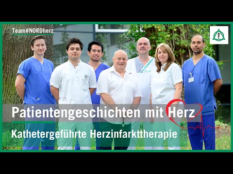
In this detailed patient recovery story, we examine the remarkable journey of Mr. Weber, a 62-year-old patient who suffered a massive heart attack—frequently referred to as the widowmaker—while spending time with his family in Germany. Rushed to Asklepios Klinik Nord in Hamburg under critical conditions requiring mechanical ventilation and temporary circulatory support, his case highlights the life-saving capabilities of advanced cardiac care in Hamburg Germany. Through expert interventions by senior physicians like Dr. Reimold, structured rehabilitation protocols, and state-of-the-art device therapy, this narrative illustrates how comprehensive Heart Attack Treatment in Hamburg Germany restores hope, health, and future tomorrows.
Video Chapters & Quick Navigation
Recognizing the Subtle and Deceptive Early Signs of Cardiac Arrest
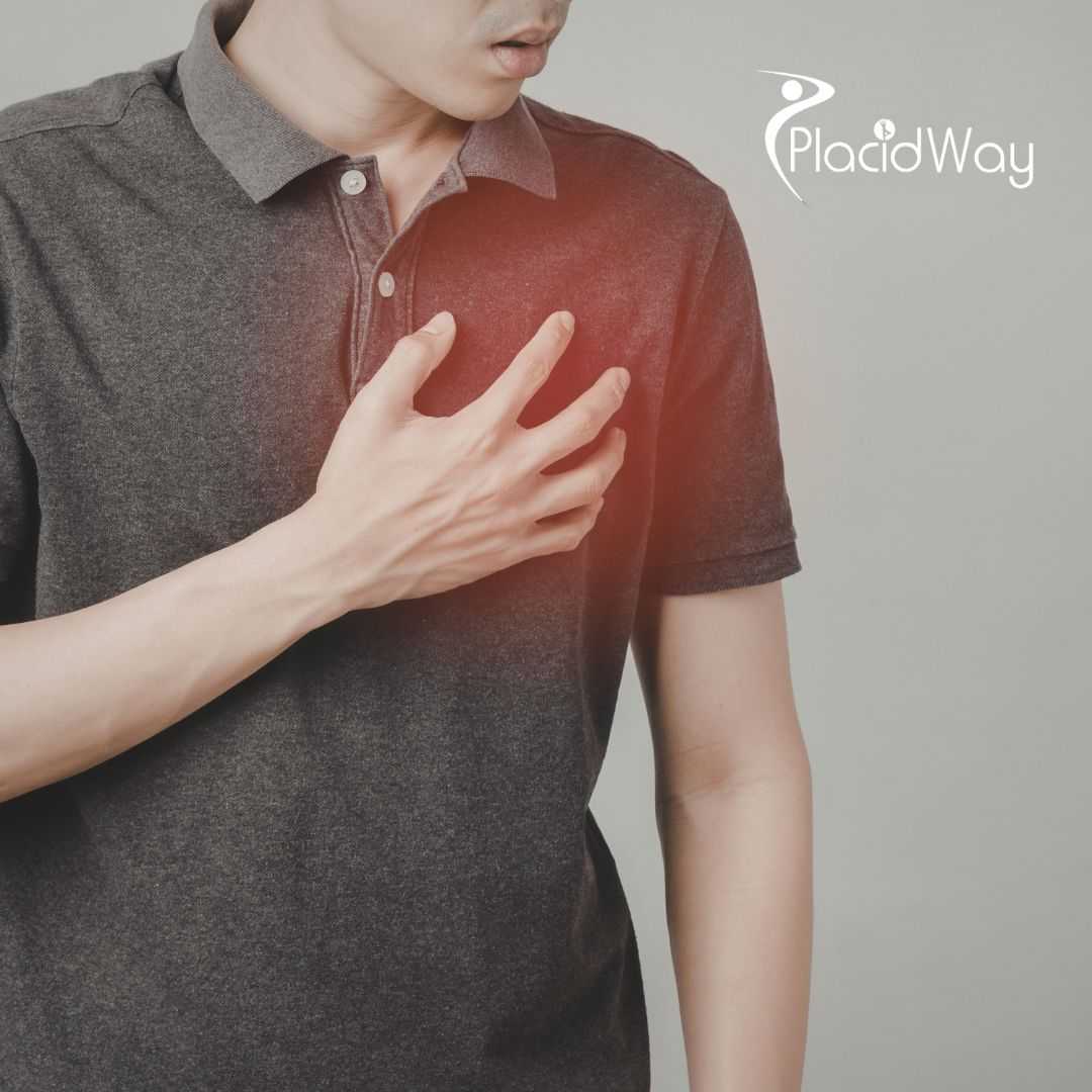
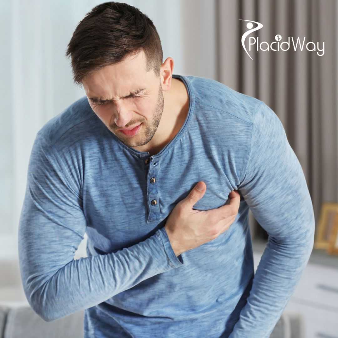
Myocardial infarctions rarely announce themselves with the dramatic clarity often depicted in cinematic media. Instead, symptoms can begin insidiously, masquerading as routine discomfort, indigestion, or fatigue. For 62-year-old Mr. Weber, the evening started innocently enough with family gardening followed by a fish dinner. However, as evening turned into night, an underlying cardiac event began unfolding silently within his coronary arteries.
As detailed in the patient interview at [00:44], Mr. Weber initially mistook his growing physical discomfort for food poisoning related to his dinner. Unable to sleep, he spent hours walking restlessly across different floors of his home, hoping movement would alleviate the sensation. It was only when crushing chest pressure intensified, accompanied by heavy cold sweating and radiating discomfort in his left arm, that the true gravity of the situation became apparent. His son-in-law's prompt realization and emergency call ultimately saved his life.
Understanding Key Cardiovascular Risk Factors and Demographics
Medical statistics consistently demonstrate that certain biological and lifestyle characteristics significantly elevate an individual's susceptibility to acute coronary syndromes. Identifying these parameters allows physicians to stratify patient risk and implement proactive preventive measures long before an emergency strikes.
As discussed during the clinical exchange at [03:14], Mr. Weber possessed multiple classic risk markers that contributed to his medical emergency. He acknowledged being a heavy smoker for decades and carrying significant excess body weight over a 20-to-30-year period. Combined with being a 62-year-old male, his physiological profile placed him squarely in a high-risk category for ischemic heart disease.
Primary Cardiac Risk Factors to Monitor Closely
- Chronic Tobacco Use: Long-term smoking damages arterial linings, accelerates plaque accumulation, and increases blood clot risks.
- Advanced Age and Gender: Men over the age of 45 face a statistically higher baseline probability of early coronary artery disease.
- Obesity and Excess Weight: Carrying excessive body weight places chronic strain on ventricular pumping function and elevates blood pressure.
- Sedentary Lifestyle: Lack of regular physical activity diminishes cardiovascular endurance and impairs healthy metabolic regulation.
- Unmanaged Dietary Habits: Diets high in sodium, trans fats, and processed sugars contribute directly to systemic arterial inflammation.
Pre-Hospital Emergency Care and Rapid Hospital Admission Protocols
When an acute myocardial infarction occurs, every minute of delayed medical intervention results in progressive and irreversible death of heart muscle tissue. The transition from a domestic emergency to intensive hospital care requires seamless coordination between emergency medical services and specialized clinical intake teams.
As explained by Dr. Reimold at [03:57], emergency medical technicians at the scene immediately identified life-threatening abnormalities using a pre-hospital electrocardiogram (ECG). However, Mr. Weber's condition rapidly deteriorated during transit due to acute respiratory distress, necessitating mechanical ventilation. He arrived at the clinic unconscious in a medically induced coma, prompting immediate echocardiography that confirmed severely impaired ventricular function and triggered urgent catheterization procedures.
| Emergency Phase | Clinical Action Performed | Patient Status & Objective |
|---|---|---|
| Pre-Hospital Assessment | On-site ECG recording by paramedics. | Rapid identification of ST-elevation myocardial infarction. |
| Emergency Transit | Mechanical ventilation and life support. | Stabilization of breathing during cardiogenic compromise. |
| Clinical Intake | Ultrasound and urgent catheterization. | Assessment of left ventricular damage and vessel blockage. |
Advanced Minimally Invasive Cardiac Catheterization and Stent Placement
Modern interventional cardiology has revolutionized the management of coronary artery blockages, replacing highly invasive open-heart surgeries with precise, catheter-based procedures performed under local anesthesia. This technological evolution drastically reduces recovery times and improves survival rates.
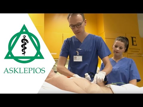
As detailed by the interventional specialist at [04:52], cardiac catheterization is a diagnostic and therapeutic X-ray procedure where a thin catheter is introduced through an artery in the wrist or groin. Contrast dye highlights blood vessel pathways, allowing physicians to pinpoint obstructions instantly. Using microscopic guide wires, miniature balloons are inflated inside the narrowed artery, followed by the deployment of permanent metal mesh tubes known as stents to maintain open blood flow.
Key Steps in Modern Percutaneous Coronary Intervention
- Vascular Access: Insertion of a fine catheter sheath into radial or femoral arteries under local anesthesia.
- Angiographic Mapping: Injection of radiopaque contrast dye to visualize coronary blood flow and blockage locations.
- Balloon Angioplasty: Inflation of a specialized micro-balloon across the obstruction to compress plaque against arterial walls.
- Stent Deployment: Permanent placement of expandable metal mesh scaffolding to keep the vessel patent.
- Post-Procedure Verification: Final contrast imaging to confirm restoration of normal myocardial perfusion.
Overcoming Cardiogenic Shock Caused by Widowmaker Artery Blockages
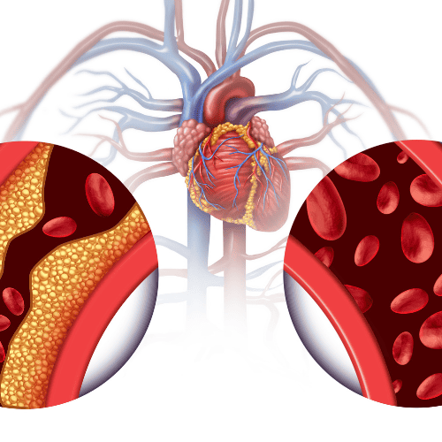
Certain coronary occlusions carry an exceptionally high mortality rate due to the sheer volume of heart muscle tissue dependent on that specific vessel. When the primary artery supplying the left ventricle becomes completely obstructed, the heart can fail to pump blood effectively, triggering cardiogenic shock.
As noted at [06:16], Mr. Weber suffered a complete occlusion in one of his three major coronary arteries—specifically the anterior descending branch often colloquially termed the widowmaker. Because the blockage occurred near its origin, blood flow to almost the entire left ventricle was interrupted. To manage this critical emergency, the clinical team utilized a temporary mechanical circulatory support device to relieve ventricular strain while restoring coronary perfusion.
The Therapeutic Role of Wearable Cardioverter-Defibrillator Vests
Following a severe myocardial infarction, patients remain at an elevated risk of sudden cardiac death due to lethal ventricular arrhythmias while their heart muscle attempts to heal. Implanting a permanent device immediately is often unnecessary because myocardial function frequently improves over time with optimized medication.
As explained by the electrophysiology expert at [07:30], patients with severely compromised left ventricular ejection fractions are frequently prescribed a wearable cardioverter-defibrillator vest. This specialized garment contains integrated ECG monitoring electrodes and automated shock delivery systems connected to an external battery pack. Worn continuously at home, the vest bridges the vulnerable healing window by detecting life-threatening rhythm disorders like ventricular fibrillation and delivering immediate life-saving shocks before emergency services can arrive.
Essential Functions of Wearable Defibrillator Vests
- Continuous Rhythm Monitoring: Multi-lead ECG sensors track heart electrical activity 24 hours a day during daily routines.
- Automated Arrhythmia Detection: Advanced algorithms instantly recognize dangerous tachycardias and ventricular fibrillations.
- Patient Alert Response: Audible and vibrational alarms allow conscious patients to cancel false alarms before shock delivery.
- Immediate Resuscitation Delivery: High-energy electrical shocks are discharged automatically to restore normal sinus rhythm.
- Safe Healing Bridge: Protects high-risk patients during the critical post-infarct recovery period prior to surgical device implantation.
Patient Reintegration, Cardiac Rehabilitation, and Lifestyle Modification
Surviving a catastrophic cardiac event is only the first phase of the healing journey; regaining physical strength and psychological confidence requires structured rehabilitation and unwavering personal commitment to lifestyle changes.
As shared by Mr. Weber during his interview at [10:25], returning to everyday life after receiving a permanent implantable cardioverter-defibrillator on June 30 involved overcoming muscle deconditioning and fatigue. Supported by his cardiologist's firm encouragement, he established a disciplined routine combining daily morning walks and afternoon exercise bike sessions. Furthermore, he successfully quit smoking permanently, embracing a healthier future dedicated to spending summers gardening with his family.
Clinical Excellence and Comprehensive Patient Support at Asklepios Kliniken
Delivering successful outcomes in complex cardiovascular emergencies demands an elite medical infrastructure, advanced diagnostic and surgical technologies, and a deeply compassionate multidisciplinary team. Asklepios Kliniken in Hamburg represents a premier European hub for specialized medical care.
Throughout Mr. Weber's journey—from emergency admission and mechanical circulatory support at Asklepios Klinik Nord in Heidberg to long-term rhythm management and rehabilitation oversight—the institution demonstrated unwavering dedication to patient welfare. By combining cutting-edge interventional cardiology with personalized follow-up care, Asklepios Kliniken continues to set benchmark standards for cardiac care in Hamburg Germany, ensuring patients receive world-class medical support when it matters most.
Looking for Specialized Cardiology Care in Europe?
PlacidWay Medical Tourism connects international patients with top-tier hospitals and expert physicians for Cardiac Care in Hamburg Germany at Asklepios Kliniken, providing seamless access to world-class cardiovascular treatments and recovery programs.
GET A FREE QUOTEView Full Video Transcript
[00:00:00] [Music]
[00:00:12] Welcome to our patient stories from the heart, filmed at Asklepios Klinik Nord in Hamburg.
[00:00:18] A picture is worth a thousand words, but we believe it is important to tell the remarkable patient story behind this image.
[00:00:29] Joining me today are our patient, Mr. Weber, and his treating senior physician, Dr. Reimold. A very warm welcome to both of you.
[00:00:38] Hello.
[00:00:44] February 21, 2021, approximately six months ago. This is a day you will always remember. You were brought to our hospital by ambulance with the sirens on and blue lights flashing. Do you remember how your symptoms began and what happened? What symptoms did you experience?
[00:01:05] Yes, I remember it very clearly because this is not something that happens often in your life. It started when we were doing some gardening at my children's house. After finishing the work, we decided to order some food. By 8 p.m., the children had gone to bed, while we men stayed up a little longer. That was when I started feeling unwell. However, I blamed it on the food, as people often do. We had eaten fish, so I thought perhaps something had been wrong with it. I couldn't fall asleep and continued feeling uncomfortable. I started walking around the house, going from one floor to another, thinking that perhaps moving around would help. After walking around the house for about four hours, I gradually began to feel pressure in my chest. I also started sweating, with beads of sweat appearing on my head. I had also experienced pain in my left arm, but I hadn't recognized what it meant, so I didn't make the connection for quite some time. Eventually, I didn't know what else to do and called for help. My son-in-law quickly realized what might be happening and called the emergency services. That is how I remember it.
[00:02:25] May I ask how old you are?
[00:02:27] I am currently 62.
[00:02:31] So, you fit some of the typical risk characteristics: male and 62 years old. When did you first think you might be having a heart attack?
[00:02:40] It was when the pressure in my chest became noticeably stronger. Before that, I thought something was wrong with my stomach. Then I began to feel as though a tight band was wrapping around my upper body, getting tighter and tighter. At that point, I realized it could no longer simply be caused by the food I had eaten.
[00:03:09] Did it happen completely out of the blue, or had you experienced any other symptoms in the days before the gardening?
[00:03:14] I hadn't experienced any symptoms at all before that. However, I should mention my medical risk factors. I was a heavy smoker until that point, and I was also significantly overweight. These had probably been issues for the previous 20 to 30 years.
[00:03:29] Those are certainly several risk factors combined. Dr. Reimold, this sounds like an emergency call in the middle of the night. How should we imagine the situation? You are an interventional cardiologist, and a 62-year-old father arrives at the hospital with breathing difficulties and in critical condition. What needs to happen next, and how is a heart attack diagnosed?
[00:03:57] In Mr. Weber's case, the diagnosis was relatively clear. The emergency physician had already identified it at the scene using an electrocardiogram, or ECG. The ECG showed clear abnormalities. Together with the symptoms Mr. Weber described, this led the emergency team to notify our hospital that they were bringing in a suspected heart attack patient. Unfortunately, his condition became more complicated on the way to the hospital because he developed additional breathing difficulties. At one point, he even required mechanical ventilation. He therefore arrived unconscious, essentially in a medically induced coma, and was being ventilated by a machine. At the hospital, we performed another assessment, including an ultrasound examination of his heart. This showed that his heart function was severely impaired. Combined with the ECG findings, this prompted us to arrange an immediate cardiac catheterization.
[00:04:45] Cardiac catheterization sounds like a major operation. What does it involve, what do you do, and what is it used for?
[00:04:52] Essentially, it is primarily an X-ray examination that allows us to visualize the blood supply to the heart muscle. During an acute heart attack, a blood vessel can suddenly become blocked, interrupting the blood supply to the heart muscle. As a result, the affected part of the heart can no longer function properly. To treat the problem, we first need to identify which blood vessel or vessels are affected. That is why we perform a cardiac catheterization. It is a minimally invasive procedure, not an operation. The procedure is performed through the skin. We insert a thin plastic tube, called a catheter, into an artery at the wrist or groin and guide it through the blood vessel against the direction of blood flow. This is generally performed under local anesthesia. We can then inject contrast dye through the catheter, allowing us to see the blood vessels on X-ray images and identify exactly where the problem is. The major advantage is that we can also treat the narrowing or blockage during the same procedure. To do this, we pass a thin wire through the catheter and into the blocked vessel. A small balloon is then guided over the wire and inflated to widen the vessel. Finally, we insert what is called a stent to keep the vessel open. A stent is a small metal mesh tube that remains inside the blood vessel and acts as a supporting framework.
[00:06:16] In Mr. Weber's case, however, we were dealing with a very severe condition. One of his three coronary arteries was completely blocked. It was the major artery supplying the front wall of the heart, and the blockage was located relatively close to its origin. This meant that the blood supply to almost the entire left ventricle was temporarily interrupted. As a result, he developed a critical condition known as cardiogenic shock. Among interventional cardiologists who perform stent procedures, this particular artery is sometimes referred to as the "widowmaker" because a blockage can be extremely dangerous. That is why we also decided to use a temporary mechanical heart-support device to relieve the heart during this critical phase while treatment was being carried out to restore blood flow.
[00:07:10] You are not only an interventional cardiologist but also an expert in defibrillator treatment at Asklepios Klinik Nord in Heidberg. Mr. Weber's heart function is now being supported with medication. But which patients need a defibrillator after a heart attack, and which patients do not?
[00:07:30] Generally, we do not implant a defibrillator immediately after a heart attack because we know that the heart muscle has some potential to recover and that heart function can improve. We also know that people with a certain degree of heart failure have an increased risk of what is known as sudden cardiac death. This means that a life-threatening heart rhythm disturbance can occur unexpectedly. One example is ventricular fibrillation, a serious rhythm disorder that can effectively cause cardiac arrest. Patients who have this risk immediately after a heart attack because of severely reduced function of the left ventricle, as was the case with Mr. Weber, may initially be given what is called a wearable cardioverter-defibrillator. Instead of implanting a device immediately, we provide the patient with a special vest containing defibrillator electrodes connected to a battery that must be worn with the device. This vest serves a similar purpose to an implanted defibrillator. It can detect life-threatening heart rhythm disturbances and deliver an electric shock immediately when necessary. In an emergency, a paramedic or emergency physician might otherwise need to arrive and administer treatment, potentially 10 to 15 minutes later. By then, it could be too late. That is why we prescribed this vest for Mr. Weber. He went home wearing it and managed to adapt to it quite well. Of course, it does impose some limitations on daily life. Patients should wear it as much as possible and for as long as recommended, ideally continuously. During this period, we want to give the medication time to reduce the strain on the heart and allow the heart muscle, particularly its pumping function, to recover. Regular ultrasound examinations then help us assess whether the heart function has improved sufficiently. In the best-case scenario, the risk of sudden, dangerous heart rhythm disturbances becomes low enough that an implanted defibrillator is no longer necessary. In Mr. Weber's case, however, even after cardiac rehabilitation and optimization of his medication, his physical exercise capacity improved, but his heart's pumping function did not recover above the threshold considered sufficiently safe. We also found that the wearable defibrillator, which functions somewhat like an ECG monitor, had recorded repeated brief episodes of potentially dangerous heart rhythm disturbances. Consequently, we decided to implant a defibrillator in Mr. Weber on June 30.
[00:10:25] That was fascinating. Thank you very much, Dr. Reimold. Mr. Weber, you now have a specialized cardiac device. You mentioned that you are an active family man who enjoys gardening and spending time with your family. How should we imagine your recovery? After suffering such a serious heart attack, you went through rehabilitation and wore a defibrillator vest. Now, after approximately six months, you have returned from the hospital with an implanted defibrillator. Are you able to participate fully in everyday life again? How is your reintegration into normal activities progressing?
[00:10:57] I am still in the middle of the recovery process. Since the implant was placed only three weeks ago, I am still experiencing some problems with my chest muscles. For example, I wouldn't lift a large crate of bottled water just yet. Unfortunately, because my movement has been restricted recently, and generally over a longer period, my muscles have also lost some of their strength. I wouldn't say they have wasted away, but they certainly don't perform as well as they used to. For example, I recently tried to go for a walk. The weather was warm, but I had to stop every 50 meters because I simply didn't have enough strength to keep going. Otherwise, I feel quite well at the moment.
[00:11:37] Do you feel that your everyday life is improving, particularly when you compare your current condition with your time in intensive care and on the general ward? Are you getting better from week to week?
[00:11:52] Yes, I can definitely say that my condition is improving. If my own tendency to put things off hadn't held me back, I would have started exercising earlier. My cardiologist here has given me a telling-off. She keeps telling me to go for walks, go for walks, and go for walks. That is the first step. I have also bought an exercise bike, which I now use every day. I go for a walk in the morning and ride the bike in the afternoon. I hope that everything will gradually get a little better.
[00:12:28] We certainly hope so, too. We want to encourage you, and your story also encourages us. We follow many patients over the years, and we see them make good progress. It is important for us to hear directly from patients how they are doing after experiencing such a severe state of shock and requiring a temporary mechanical heart-support device, like the one we implanted and subsequently removed at Asklepios Klinik Nord in Heidberg to support you through that critical period. From what we can see, you really do seem to be moving in the right direction, one small step at a time.
[00:13:15] Absolutely. That makes me very happy. Next week—sorry, next week I have... And, of course, I have given up smoking...
[00:13:26] I didn't mean to interrupt you. I'm sorry. My colleagues are naturally delighted that you are doing well. On behalf of the entire heart team at Asklepios Klinik Nord, we would also like to send you our warmest regards. We are very pleased that you are feeling better. If you have any questions or need any further assistance, we are here for you and your cardiologist. We are delighted to see your improvement and wish you continued recovery.
[00:13:48] Thank you. I certainly hope so, too. I hope to spend many more summers gardening with my children and their spouses.
[00:14:01] On behalf of Dr. Reimold as well, thank you very much for agreeing to take part in this interview. And thank you to everyone watching for your interest in these patient stories from the heart at Asklepios Klinik Nord in Heidberg.
[00:14:18] Thank you very much.
[00:14:19] [Music]














Share this listing