


Request a video call
Confirm Your Virtual Consultation
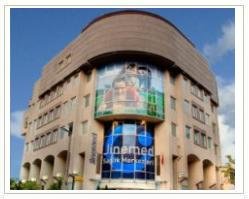
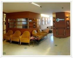
Jinemed Hospital | Orthopedic Clinic Profile Overview
-
Accurate diagnosis and effective prompt treatment.
-
We want to relieve pain, restore the function of an organ or of the body and bring your health to an upper level.
-
Come and enjoy the highest level of service provided by our dedicated specialists. We are ready to give you what you deserve and extend our service beyond your expectations.
-
We are always happy to answer any questions you might have about health related issues, the staff, the treatment options, the payment options. Do not forget whatever your age is one day you might need physiotherapy.
Orthopedic Surgery is one of the most demanded fields at Jinemed Hospital and we will be happy to assist our patients regarding these surgeries. We offer procedures from knee replacements to hip replacements, from arthroscopic surgeries to shoulder surgeries. Jinemed Hospital has long developed its reputation as a world-class and well-known facility focusing in orthopedic surgery for individuals in Central Europe, Asia, and as far away as America. Located in Istanbul, Turkey, Jinemed Hospital has well experienced team that specializes in all types of orthopedic surgeries such as:
-
Knee Replacement Surgery
-
Hip Replacement Surgery
-
Arthroscopic Meniscal
-
Cartilage Surgery
-
Knee ACL
-
Rotator Cufff Tear
-
Shoulder Imp. Syndrome
-
S.L.A.P. Tear
-
And Alot more...
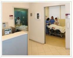
JInemed Hospital is one of the most technologically advanced medical center for orthopedic surgeries, and offers excellent, experienced an affordable orthopedic care for knee injuries, sports injuries and back and spine care.
Orthopedic surgeries are performed on bones and joints. Such surgeries offer a variety of treatments and procedures that restore range of motion, flexibility, and enhanced quality of life for individuals suffering from arthritis, rheumatism, sports injuries, auto accidents, and disease processes that destroy connective tissues, bones and joints.
Our Surgeon:
Dr. Selim M. is a member of İstanbul Bakırköy Research and Training Hospital Orthopeadics and Traumatology Service (As a fellowship in Trauma) and a well experienced and highly qualified surgeon. He specializes in all types of orthopedic surgeries including ankle and foot surgeries and trauma surgeries.
The Jinemed Hospital is located in the middle of the city on the eastern side of Istanbul, where international patients receive world-class medical service, a modern facility, and the benefit of English speaking personnel.
Our locations:
We are located at the European side of İstanbul: FULYA SPORTOMED.
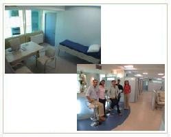
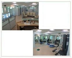
Fulya Sportomed is located at the intersection of some main avenues of the city: Hakki Yeten Avenue No:19, Floor:4 . İt is placed on a surface of 400 squaremeters. We can give service to 18 people at the same time. We provide cabines with phone, internet service, lockers and showers. Our customers can enjoy a relaxation at the special sections and they can also enjoy drinks at ´the vitamin bar´.
We have all kind of physical therapy equipment, exercises and rehabilitation equipment. We have isokinetique exercise and test equipment CYBEX. Physical Therapy Specialist Doctors, A Sport Medicine Specialist Doctor and Physiotherapists are on duty.
Our second location is at the Asian side of the city on the most attractive avenue in İstanbul: SUADİYE SPORTOMED. Bagdat Caddesi No 471. İt covers a surface of 300 squaremeters and 18 patients can be provided service at the same time. We have physical therapy equipment, exercises and rehabilitation equipment and CYBEX testing and exercise system. Physical Therapy Specialist Doctors, A Sport Medecine Specialist Doctor and Physiotherapists are on duty.
Please Click here to request more information from Jinemed Hospital Medical Center.
Jinemed Hospital | Orthopedic Clinic, Istanbul, Turkey Profile Details
OUR VALUES
Our main values are based on 2 essential principles: general work ethics and patients rights.
Our general work principles:
- The main aim of the therapy is to help the patient reach the quality of life s/he had before the disease or injury.
- We respect the patient´s right unconditionally.
- Each person is treated as an individual. We apply holistic approach and try to diagnose all medical problems.
- We respect a patient´s convenience and we try to arrange the therapy time. We provide flexible appointments.
- We respect a person´s right to have information during the therapy sessions: we provide the individual with the details of treatment regime, its benefits and risks and we offer choices; eventually we respect any individual choice.
- We provide our clients with a clean an hygienic treatment place.
- Our patients know that there are therapists and a doctor on site.
- We follow the medical and technical advance in our field.
- We want to give a high standard service.
Patient rights:
- We know that each patient has a right to have a healthy life and has a right to be provided by new treatment options.
- We offer equal service to individual regardless of his/her age, sex, religion, ethnic group.
- We give medical information to our patients.
- We respect a person´s right to start and terminate treatment and to choose his/her medical team and to change service provider.
- We respect the right to ask the details of the treatment offered.
- Confidentiality is essential during the offer of a service.
- We always ask for consent before starting a treatment.
- We offer our clients a clean and secure environment.
- Everyone has a right to be treated respectfully.
- Our patients have the right to accept visitors and likewise they have right to be accompanied by a third person during the examination and the treatment.
- The customer has a right to make complaints.
- The customer has a right to have ongoing service.
Hotel Accommodation
IVF Turkey offers both economic hotels, and luxury hotels in Istanbul.
Some of the hotels we work with are the following :
- Ta?l?k Hotel
Please Click here to request more information from Jinemed Hospital Medical Center.
Jinemed Hospital | Orthopedic Clinic Treatments Offered
Prices:
| PROCEDURES | Stay | PRICE in GBP |
| ACL Repair | 2 | 3750 |
| Ankle fusion | 3 | 2500 |
| Ankle Joint Fusion | 3 | 3750 |
| Ankle Joint Replacement | 3 | 7500 |
| Arthroscopy Elbow | 3 | 1875 |
| Arthroscopy Knee | 3 | 1875 |
| Arthroscopy Menisectomy | 3 | 2188 |
| Arthroscopy Shoulder | 3 | 3125 |
| Subtolar Joint | 3 | 1875 |
| CTS | 3 | 1250 |
| Dupuytren | 3 | 2500 |
| EpicondylitisWilhelm | 3 | 1875 |
| Finger or Toe Endoprothesis | 3 | 2500 |
| Hallux Valgus | 3 | 3125 |
| Hammer Toe | 3 | 1250 |
| Hip Replacement Minimum Invasive | 10 | 7500 |
| Hip Replacement Primary | 10 | 7500 |
| Hip Resurfacing | 10 | 7500 |
| Knee Replacement Primary | 10 | 8750 |
| Knee Replacement Revision | 10 | 11250 |
| Knee Replacement Unicondylar | 10 | 8750 |
| Shoulder Endoprosthesis Partial | 10 | 7500 |
| Shoulder Endoprosthesis Total | 10 | 8750 |
Orthopaedics–Traumatology:
Various problems from a simple injury to complex fractures incurred after trauma are treated with modern methods. Jinemed has special clinics in Foot Disease and Back Disease.
Surgeries include:
Arthroscopic Meniscal Surgery
What is arthroscopic meniscal surgery?
Arthroscopic meniscal surgery is a procedure in which a surgeon uses an arthroscope and other tools to remove all or part of a damaged meniscus in the knee or, if possible, to repair a meniscus. A meniscus is a piece of rubbery tissue (fibrocartilage) between the bones of the knee joint. An arthroscope is a tube with a light at the end that projects an image of the inside of your knee onto a TV monitor. The arthroscope is about the diameter of a pencil.
When is it used?
The procedure is used when you have damaged cartilage in your knee.
Examples of alternatives are:
- limiting your activity
- taking medicine to reduce the swelling
- having physical therapy
- having open knee surgery
- choosing not to have treatment, while recognizing the risks of your condition.
You should ask your health care provider about these choices.
How do I prepare for this procedure?
Plan for your care and recovery after the operation, especially if you are to have general anesthesia. Allow for time to rest and try to find other people to help you with your day-to-day duties.
Follow instructions provided by your health care provider. Do not eat or drink anything after midnight or the morning before the procedure. Do not even drink coffee, tea, or water.
What happens during the procedure?
You will be given a general, regional, or local anesthetic. A general anesthetic will relax your muscles and make you feel as if you are in a deep sleep. Both local and regional anesthetics numb part of the body while you remain awake. All three types of anesthesia should keep you from feeling pain.
The surgeon will put an arthroscope and one or two tools into the knee joint through small cuts. Fluid is injected into the knee to expand the joint so that the structures and cartilage can be seen. The surgeon will examine the knee to find any damage. She or he may repair any torn cartilage or shave down the cartilage in the knee and remove the pieces of cartilage. The surgeon will then remove the arthroscope and the tools and close the small openings with stitches.
What happens after the procedure?
You will go home the same day. You should keep your leg elevated. Take it easy for at least the next 2 to 3 days. Do not take part in strenuous activities until your health care provider feels you are ready.
After surgery:
- Use crutches for several days or until you can walk nearly normally.
- Elevate your leg so that your ankle is higher than your knee and your knee is higher than your hip.
- Put ice on your knee for 20 to 30 minutes 3 or 4 times a day until symptoms are gone.
- Start bending your knee as soon as possible.
- Change your bandage after 4 days and cover the cuts with band-aids or gauze.
- If you have a brace or splint, consult your health care provider.
- If the cartilage is repaired and not trimmed, we want you to use crutches longer and to not put weight on your leg.
What are the benefits of this procedure?
The arthroscopy may treat the knee without the need for open knee surgery with bigger incisions. There is more rapid recovery than with open knee surgery.
What are the risks associated with this procedure?
- There are some risks when you have general anesthesia. Discuss these risks with your health care provider.
- The nerves around the knee may be injured causing a little spot of numbness in the leg below the knee.
- There is a risk of deep vein thrombosis, a condition in which a blood clot forms within a deep-lying vein.
- There is a risk of infection and bleeding.
Cartilage Surgery
What are the signs and symptoms of an articular cartilage injury that may be treated with the microfracture technique?
- Intermittent swelling - Loose fragments floating in the knee can cause swelling to occur.
- Pain - Pain with prolonged walking or climbing stairs can occur.
- Giving way - The knee may occasionally buckle or give way when weight is placed upon it.
- Locking or catching - Loose, floating pieces of cartilage may catch in the joint as it bends, causing the knee to lock or have limited motion.
- Noise - The knee may make noise (called crepitus) during motion, especially if the cartilage on the back of the kneecap is damaged. This noise is often described as "snap, crackle, and pop".
The microfracture procedure is done arthroscopically. The surgeon visually assesses the defect and performs the procedure using special instruments that are inserted through three small incisions on the knee. After assessing the cartilage damage, any unstable cartilage is removed from the exposed bone. The surrounding rim of remaining articular cartilage is also checked for loose or marginally attached cartilage. This loose cartilage is also removed so that there is a stable edge of cartilage surrounding the defect. The process of thoroughly cleaning and preparing the defect is essential for optimum results.
Multiple holes, or microfractures, are then made in the exposed bone about 3 to 4mm apart. Bone marrow cells and blood from the holes combine to form a "super clot" that completely covers the damaged area. This marrow-rich clot is the basis for the new tissue formation. The microfracture technique produces a rough bone surface that the clot adheres to more easily. This clot eventually matures into firm repair tissue that becomes smooth and durable. Since this maturing process is gradual, it usually takes two to six months after the procedure for the patient to experience improvement in the pain and function of the knee. Improvement is likely to continue for about 2 to 3 years.
Rehabilitation
- Rehabilitation Protocol for Patients with Chondral Defects on the Femur or Tibia.
- The patient begins passive flexion/extension (straightening and bending) of the knee with 500 repetitions three times a day.
- The use of crutches, with only light touch-down weight allowed on the involved leg, is prescribed for 6 to 8 weeks.
- Patients with small defect areas (less than 1cm in diameter) may be allowed to put weight on the leg a few weeks sooner.
- Limited strength training also begins immediately after microfracture surgery.
- Standing one-third knee bends with a great deal of the weight on the uninjured leg begin the day after surgery.
- Stationary biking without resistance and a deep-water exercise program begin 1 to 2 weeks after surgery.
- After 8 weeks the patient progresses to full weight bearing and begins a more vigorous program of active knee motion.
- Elastic resistance cord exercises can begin about 8 weeks following surgery.
- Free weights or machine weights can be started when the early goals of the rehabilitation program have been met, but no sooner than 16 weeks after surgery.
- Patients must not resume sports that involve pivoting, cutting, and jumping for 4 to 6 months after a microfracture procedure.
- Full activity may be resumed once the physician has examined the knee and given approval for the patient to return to sports activity.
Knee ACL
Non-Operative Treatment Benefits and Limits
Surgical treatment is usually advised in dealing with combined injuries (ACL tears in combination with other injuries in the knee). However, deciding against surgery is reasonable for select patients. Non-operative management of isolated ACL tears is likely to be successful or may be indicated in patients:
- With partial tears and no instability symptoms
- With complete tears and no symptoms of knee instability during low-demand sports who are willing to give up high-demand sports
- Who do light manual work or live sedentary lifestyles
- Whose growth plates are still open (children)
Surgical Intervention and Considerations
Patients treated with surgical reconstruction of the ACL have long-term success rates of 82 percent to 95 percent. Recurrent instability and graft failure are seen in approximately 8 percent of patients. The goal of the ACL reconstruction surgery is to prevent instability and restore the function of the torn ligament, creating a stable knee. This allows the patient to return to sports. There are certain factors that the patient must consider when deciding for or against ACL surgery.
Patient considerations
Active patients involved in sports or jobs that require pivoting, turning or hard-cutting as well as heavy manual work are encouraged to consider surgical treatment. This includes older patients who have previously been excluded from consideration for ACL surgery. Activity, not age, should determine if surgical intervention should be considered.
In young children or adolescents with ACL tears, early ACL reconstruction creates a possible risk of growth plate injury, leading to bone growth problems. The surgeon can delay ACL surgery until the child is closer to skeletal maturity or the surgeon may modify the ACL surgery technique to decrease the risk of growth plate injury.
A patient with a torn ACL and significant functional instability has a high risk of developing secondary knee damage and should therefore consider ACL reconstruction.
It is common to see ACL injuries combined with damage to the menisci (50 percent), articular cartilage (30 percent), collateral ligaments (30 percent), joint capsule, or a combination of the above. The "unhappy triad", frequently seen in football players and skiers, consists of injuries to the ACL, the MCL and the medial meniscus. In cases of combined injuries, surgical treatment may be warranted and generally produces better outcomes. As many as 50 percent of meniscus tears may be repairable and may heal better if the repair is done in combination with the ACL reconstruction.
Surgical choices
The middle third of the patellar tendon of the patient, along with a bone plug from the shin and the knee cap is used in the patellar tendon autograft . Occasionally referred to by some surgeons as the "gold standard" for ACL reconstruction, it is often recommended for high-demand athletes and patients whose jobs do not require a significant amount of kneeling. In studies comparing outcomes of patellar tendon and hamstring autograft ACL reconstruction, the rate of graft failure was lower in the patellar tendon group (1.9 percent versus 4.9 percent). In addition, most studies show equal or better outcomes in terms of postoperative tests for knee laxity (Lachman's, anterior drawer and instrumented tests) when this graft is compared to others. However, patellar tendon autografts have a greater incidence of postoperative patellofemoral pain (pain behind the kneecap) complaints and other problems. The pitfalls of the patellar tendon autograft are:
- Postoperative pain behind the kneecap
- Pain with kneeling
- Slightly increased risk of postoperative stiffness
- Low risk of patella fracture
The semitendinosus hamstring tendon on the inner side of the knee is used in creating the hamstring tendon autograft for ACL Reconstruction. Some surgeons use an additional tendon, the gracilis, which is attached below the knee in the same area. This creates a two- or four-strand tendon graft (Figure 8). Hamstring graft proponents claim there are fewer problems associated with harvesting of the graft compared to the patellar tendon autograft including:
- Fewer problems with anterior knee pain or kneecap pain after surgery
- Less postoperative stiffness problems
- Smaller incision
- Faster recovery
The graft function may be limited by the strength and type of fixation in the bone tunnels, as the graft does not have bone plugs. There have been conflicting results in research studies as to whether hamstring grafts are slightly more susceptible to graft elongation (stretching), which may lead to increased laxity during objective testing. Recently, some studies have demonstrated decreased hamstring strength in patients after surgery.
The quadriceps tendon autograft is often used for patients who have already failed ACL reconstruction. The middle third of the patient's quadriceps tendon and a bone plug from the upper end of the knee cap are used. This yields a larger graft for taller and heavier patients. Because there is a bone plug on one side only, the fixation is not as solid as for the patellar tendon graft. There is a high association with postoperative anterior knee pain and a low risk of patella fracture. Patients may find the incision is not cosmetically appealing.
Allografts are grafts taken from cadavers (Figure 10) and are becoming increasingly popular. These grafts are also used for patients who have failed ACL reconstruction before and in surgery to repair or reconstruct more than one knee ligament. Advantages of using allograft tissue include elimination of pain caused by obtaining the graft from the patient, decreased surgery time and smaller incisions. The patellar tendon allograft allows for strong bony fixation in the tibial and femoral bone tunnels with screws. However, allografts are associated with a risk of viral transmission, including HIV and Hepatitis C, despite careful screening and processing. Several deaths linked to bacterial infection from allograft tissue (due to improper procurement and sterilization techniques) have led to improvements in allograft tissue testing and processing techniques. There have also been conflicting results in research studies as to whether allografts are slightly more susceptible to graft elongation (stretching), which may lead to increased laxity during testing.
Operative Procedure
Before any surgical treatment, the patient is usually sent to physical therapy. Patients who have a stiff, swollen knee lacking full range of motion at the time of ACL surgery may have significant problems regaining their motion after surgery. It usually takes three or more weeks from the time of injury to achieve full range of motion. It is also recommended that some ligament injuries be braced and allowed to heal prior to ACL surgery. The patient, the surgeon and the anesthesiologist select the anesthesia used for surgery. Patients may benefit from an anesthetic block of the nerves of the leg to decrease postoperative pain. The surgery usually begins with an examination of the patient's knee while the patient is relaxed due the effects of anesthesia. This final examination is used to verify that the ACL is torn and also to check for looseness of other knee ligaments that may need to be repaired during surgery or addressed postoperatively. If the physical exam strongly suggests the ACL is torn, the selected tendon is harvested (for an autograft) or thawed (for an allograft) and the graft is prepared to the correct size for the patient. After the graft has been prepared, the surgeon places an arthroscope into the joint. Small (one-centimeter) incisions called portals are made in the front of the knee to insert the arthroscope and instruments and the surgeon examines the condition of the knee. Meniscus and cartilage injuries are trimmed or repaired and the torn ACL stump is then removed. In the most common ACL reconstruction technique, bone tunnels are drilled into the tibia and the femur to place the ACL graft in almost the same position as the torn ACL. A long needle is then passed through the tunnel of the tibia, up through the femoral tunnel, and then out through the skin of the thigh. The sutures of the graft are placed through the eye of the needle and the graft is pulled into position up through the tibial tunnel and then up into the femoral tunnel . The graft is held under tension as it is fixed in place using interference screws, spiked washers, posts or staples. The devices used to hold the graft in place are generally not removed. Variations on this surgical technique include the "two-incision" and "over-the-top" types of ACL reconstructions, which may be used because of the preference of the surgeon or special circumstances (revision ACL reconstruction, open growth plates). Before the surgery is complete, the surgeon will probe the graft to make sure it has good tension, verify that the knee has full range of motion and perform tests such as the Lachman's test to assess graft stability. The skin is closed and dressings (and perhaps a postoperative brace and cold therapy device, depending on surgeon preference) are applied. The patient will usually go home on the same day of the surgery.
Potential Operative Complications
The incidence of infection after arthroscopic ACL reconstruction has a reported range of 0.2 percent to 0.48 percent. There have also been several reported deaths linked to bacterial infection from allograft tissue due to improper procurement and sterilization techniques. Allografts specifically are associated with risk of viral transmission, including HIV and Hepatitis C, despite careful screening and processing. The chance of obtaining a bone allograft from an HIV-infected donor is calculated to be less than 1 in a million. We do not use allografts at our hospital. Rare risks include bleeding from acute injury to the popliteal artery (overall incidence is 0.01 percent) and weakness or paralysis of the leg or foot. It is not uncommon to have numbness of the outer part of the upper leg next to the incision, which may be temporary or permanent. A blood clot in the veins of the calf or thigh is a potentially life-threatening complication. A blood clot may break off in the bloodstream and travel to the lungs, causing pulmonary embolism or to the brain, causing stroke. This risk of deep vein thrombosis is reported to be approximately 0.12 percent. Recurrent instability due to rupture or stretching of the reconstructed ligament or poor surgical technique (reported to be as low as 2.5 percent and as high as 10 percent) is possible. Knee stiffness or loss of motion has been reported at between 5 percent and 25 percent. Rupture of the patellar tendon (patellar tendon autograft) or patella fracture (patellar tendon or quadriceps tendon autografts) may occur due to weakening at the site of graft harvest. In young children or adolescents with ACL tears, early ACL reconstruction creates a possible risk of growth plate injury, leading to bone growth problems. The ACL surgery can be delayed until the child is closer to reaching skeletal maturity. Alternatively, the surgeon may be able to modify the technique of ACL reconstruction to decrease the risk of growth plate injury. Postoperative anterior knee pain is especially common after patellar tendon autograft ACL reconstruction. The incidence of pain behind the kneecap varies between 4 percent and 56 percent in studies, whereas the incidence of kneeling pain may be as high as 42 percent after patellar tendon autograft ACL reconstruction.
Rehabilitation/Convalescence
DAYS 1-7 POST OP
- Remain in bed with ACL-reconstructed leg elevated above heart.
- When walking (to bathroom only), put as much weight on the ACL-reconstructed leg as tolerated
- Maintain full extension on the ACL-reconstructed leg.
- Maintain at least 120° of flexion on the ACL-reconstructed leg.
- Maintain full flexion on the graft-donor leg.
- Use the shuttle to work on increasing strength on the graft-donor leg.
ONE WEEK POST OP
- Activity level will be determined by the condition of knees (activity increases as knee progresses).
full extension
- lock knee straight while standing
- heel props
- prone hangs
- towel stretches
flexion to at least 120°
- heel slides
- shuttle slides
- wall slides
decreased swelling
- use the Cryo-Cuff® (especially after exercises)
TWO WEEKS POST OP
- Activity level will be determined by the condition of knees (activity increases as knee progresses).
Maintain extension
- lock knee straight while standing
- heel props
- prone hangs
- towel stretches
Fexion should be at least 135°
- heel slides
- wall slides
- supine flexion hangs
Decrease swelling
- use Cryo-Cuff® as needed
Regaining patellar tendon strength is the main focus of rehabilitation between weeks two to four. Ideally, the patient should perform high repetitions of strengthening exercises several times a day.
Physical therapy will design an individual rehabilitation program according to the patient's objectives and goals.
FOUR WEEKS POST OP
Activity level will be determined by the condition of knees. Ability to return to activities will depend on the full range of motion and lack of swelling in the ACL-reconstructed leg.
Maintain extension
- lock knee straight while standing
- heel props
- prone hangs
- towel stretches
Full flexion
- heel slides
- supine flexion hangs
- sit back on heels
Decrease swelling
- Cryo-Cuff® use as needed
Increase strength and conditioning
- leg press - single leg
- knee extension - single leg
- step down exercise
- Stairmaster ® or bike or elliptical
Sport specific agility
- forward running
- backward running
- jump rope
- lateral slides
- crossovers
- shooting baskets, etc.
TWO, FOUR, & SIX MONTHS POST OP
Physical therapy will continue to monitor rehabilitation as the patient returns to their preoperative, fully competitive level of activity. Strength, range of motion, and swelling will be evaluated at each vistit. Rehabilitation and sporting activities will be advanced as strength, comfort, and confidence allows.
Rotator Cufff Tear
Not all rotator cuff tears require surgery.
rotatorMany patients are content with their progress following a non-operative treatment plan. Patients who have been unable to regain lost motion and strengthen the surrounding muscles sufficiently may need a rotator cuff repair. This is often the case for the younger, more active patients who want to address continued weakness following physical therapy.
Rotator cuff repairs can be performed either arthroscopically or with open incisions.
What types of complications may occur?
Complication rates after surgery are generally low. Infection tends to occur a little less often when arthroscopic techniques are used. Risks of major bleeding or nerve damage are extremely small. Postoperative stiffness is the major complication of both impingement and rotator cuff tears.
Postoperative treatment - rehabilitation
- Incisions must be kept dry for two or three days after surgery. If you have stitches they are usually removed 10 - 12 days after surgery.
Rotator cuff recovery requires more supervision. In order to achieve a full and rapid recovery, surgery should be performed as soon as the patient has full range of motion and has gained good muscle strength from a physical therapy program started when the injury is first detected. After surgery, the patient follows a closely monitored program:
- Therapy is carefully controlled in the first 6 - 12 weeks while the tendons heal back to the bone.
- The first goal is to regain full motion within 3 months after surgery. A therapist will assist in the early stages with gradual recovery of motion.
- During the first 6 weeks, there is no active use of the shoulder in order to protect the surgical repair.
- Once initial healing is achieved, a progressive stretching and strengthening program should begin.
- Full recovery can take more than 6 months. Some patients may require more time to regain muscle strength and complete the healing process.
Shoulder Impingement Syndrome
ShoulderImpingement and rotator cuff tears can be treated non-operatively or with surgery. Treatment for impingement usually begins with a non-operative treatment plan.
More than 2/3 of impingement patients can expect significant improvement in their symptoms with a physical therapy program
alone. These results are lower in older patients and in those with large bone spurs.
Non-Operative Treatment
Non-operative treatment is similar for both impingement and rotator cuff tears. A vast majority of patients improve with this primary treatment alone.
The goals of a physical therapy program include:
- strengthening the rotator cuff tendons.
- stretching and regaining lost motion caused by pain and inflammation.
- allowing the humerus to be better positioned under the acromion, thus reducing compression of the bursa.
A non-operative treatment plan is often all that is necessary for most patients with impingement syndrome. However, the small percentage of patients whose symptoms have not improved after 6 months of dedicated physical therapy may be candidates for surgery. The shoulder should be reevaluated to make sure no other problems exist.
Operative Treatment
Subacromialdecompressionexpands the space between the acromion and rotator cuff tendons. This can be done either arthroscopically or with open incisions, depending on the preference of the surgeon. During an arthroscopy, a tiny fiberoptic instrument is inserted into the joint. In many cases, the doctor can assess and repair the damage through this scope without making large incisions. Scar tissue or bone spurs can successfully be removed with either technique. If a rotator cuff tear is found at the time of surgery, it can also be repaired if necessary.
- Postoperative treatment and rehabilitation
- Incisions must be kept dry for two or three days after surgery.
Rehabilitation programs for impingement
Impingement rehabilitation begins almost immediately:
- Exercises to regain shoulder motion usually begin with a therapist in the first week after surgery and continue for about 6 weeks.
- At 6 weeks, most patients have regained full motion and will continue to regain strength with a home exercise program.
- Full recovery time after surgery varies; most patients have greatly improved at 3 months and are close to normal by 6 months.
S.L.A.P. Tear
Patients with glenoid labrum injuries present with non-specific shoulder pain that is associated with activity. External rotation and horizontal adduction are generally painful, and a click or pop may be felt with manipulation. Often there is associated weakness of the rotator cuff muscles. A very common labral injury is a tear that occurs on the top of the labrum, extending from the front to the back of the cartilage. This is known as a SLAP tear ("SLAP" is an acronym for superior labral anterior to posterior tear). This injury affects the attachment of the biceps tendon to the glenoid. An injury in this area can be extremely painful, and can cause the biceps tendon to rupture.
- slap_tearRehabilitation
- 0 - 3 weeks
- Immobilising with arm in sling day and night. (The sling should be secured to the upper body with a safety pen or use a special sling).
Movement restrictions, Passive Range Of Motion (PROM)
- 90° abduction
- 20° extension
- 70° internal rotation
- 0° external rotation
Exercises:
Swinging exercises at a minimum of 3 times a day.
Elbow and wrist does a full active range of motion (AROM) minimum 4 times a day.
3 - 6 weeks
Immobilising in the sling during the night. Remove the sling during the day and start physiotherapy.
- Movement restrictions (PROM)
- NB! Within the pain boundaries
90° abduction
20° extension
70° internal rotation
0° external rotation
Exercises:
Manual resistant movement in the scapula.
Reverse rowing, push-ups against the wall abduction in the scapulas plane (“scaption”)
External rotation, internal rotation and biceps curl.
Careful exercises both active and passive for the shoulder.
Positive body awareness training (shoulders back and the chest forward).
NB! Avoid pressure on the suture and the sutured tissue.
Goal: at the end of this period full active range of elevation
6 - 12 weeks
Immobilising in the sling is not necessary. Can be used if you need it.
No restrictions of movement.
Exercises:
Start AAROM (Assisted Active Range Of Motion)
Joint mobilising for scapula and glenohumoral mobility.
Careful exercises for the rotator cuff and the scapula stabiliser.
Increase the AROM, emphasise the humeroscapular rhythm.
Functional training: muscular control and proprioceptive exercises. Specific training with consideration to rehabilitation according to type of work or sport.
Goal: Full active range of motion.
Physical Therapy
SPORTOMEDfulya01An outpatient clinic specializing in the treatment of rheumatological, neurological and orthopedic diseases; sport related injuries and general rehabilitation.
One of the most famous and the most trusted instutition in this field in Europe.
This is a clinic established in 1997.
Since then we have succesfully completed the treatment of general public suffering from different diseases and the treatment of elite athletes and sportmen from different countries.
WHY PHYSICAL THERAPY AND REHABILITATION
fulya02When we apply physical treatment agents, for example, heat, cold or electrical currents to an individual, we can overcome pain, inflamation, muscle rigidity and muscle spasm. As a result we can obtain a quick resolution of a muscle or joint problem and we can see the related pain diminish or completely finish.
WHAT MEANS PHYSICAL THERAPY AND REHABILITATION
When we say Physical Therapy and Rehabilitation we mean the treatment of bodily function insufficiency or loss following a disease, a trauma or an injury. After physical therapy and rehabilitation sessions a person will most probably be able to accomplish his/her daily activities of living.
The most commonly used physical therapy agents are superficial heat such as infrared light or hot packs application, deep application of heat by diathermy apparatus or ultrasound machines; cold application mostly in the beginning phase of certain diseases and the application of some electrical currents.
Whereas the application of physical therapy agents is useful in the beginning of most joint and muscle problems, a following rehabilitation program provides us with more sustainable results that help a sick person to return to his/her previous quality of life. That is why rehabilitation process comes after a physical therapy program or they are applied all together.
fulya03Rehabilitation mostly consists of therapeutic exercises and it means the regain of bodily functions that are lost due to some diseases.
Firstly the meaning of rehabilitation was the treatment of invalidity. As a result of recent advance in this field, it has now a broader meaning: it covers all treatment activities by physical interventions that aim the restart of physical, psychological, social and economical independence of a sick or disabled person.
Physical Therapy and Rehabilitation is always applied by a team consisting of Spacialist Doctors, Physical Therapists and some other disciplines members such as Speech Therapists and Occupational Therapists.
The application of Physical Therapy and Rehabilitation is foreseen during the course following diseases: Pain related to rheumatological diseases; vertebrae pain: pain in the neck, dorsal region and lower back; some neurological diseases; orthopedical diseases and surgeries and sport injuries.
fulya04SPORTOMED is a physical therapy and rehabilitation instutition located in ?stanbul.
We give service to our patients in 2 different locations: one at the European side of the city the other at the Asian side of the city.
We provide all kind of Physical Therapy and Rehabilitation for the prevention and the treatment of all kind of sport injuries; for the treatment of Neurological diseases, Rheumatological diseases and Orthopedic operations, for instance, after hip, knee, ankle and shoulder oparations as well as joint replacements.
We treat neck pain, back pain, hip and groin pain, wrist and hand pain, tennis or golfer elbow, frozen shoulder, rib and chest pain, sciatica, whiplash, headaches, balance problems and very recently foot problems.
Please Click here to request more information from Jinemed Hospital Medical Center.
Jinemed Hospital | Orthopedic Clinic Certificates, Accreditations, Qualifications Treatments Offered
Orthopedic Surgeon
Dr. SELIM M.
EDUCATIONAL DATA
May 2006
Sarasota Memorial Hospital ,Florida Foot And Ankle Service with Dr.Harold Vogler
October 2004:
New York Hospital for Special Surgery Foot and Ankle Service Education for 3 weeks
September -November2003:
New York Hospital for Special Surgery Foot and Ankle Service
Education for 8 weeks
- 17 March 2002 – May 2004 : ?stanbul Bak?rköy Research and Training Hospital Orthopeadics and Traumatology Service (As a fellowship in Trauma)
- 01 December 1997- 05 March 2002 : Haydarpa?a Numune Training And Research Hospital . Orthopeadics and Traumatology Residency Program - Ankara - Turkey
- 1995 – 1997 : International Hospital. Preliminary Program which contents 6 months of emergency medicine, 6 months of ICU, 6 months of surgery.
- 1994 – 1995 : 18 months of obligatory military service as a practioner
- 1993 – 1994 : 12 months of obligatory practioner service
- 1986 – 1993 : ?stanbul University ?stanbul Medical Faculty
- 1980 – 1986 : ?stanbul Moda Collage
ATTENDED COURSES
3-7 November 1999 : 16th National Congress of Turkish Orthopeadics and Traumatology Society
1-3 November 1999 : Functional Fracture Treatment Symposium , Antalya
30 January –
04 February 2000 : Sports Medicine Of The Foot and Ankle Course Idoha, USA
3-6 April 2000 : 5 th Mediterranean Basic Arthroscopic Knee Surgery Course ,Antalya
14-16 April 2000 :?stanbul University ?stanbul Medical Faculty 2nd Deformity Course, ?stanbul
19-21 May 2000 : Complete Foot Care Course Seattle, USA
15-16 September 2000 : 4th International Foot And Ankle Surgery Course Bad Homburg, Germany
27-28 October 2000 : Diabetic Foot Course,Providence ,USA
2-5 November 2000 : Comprehensive Foot and Ankle, Arizona, USA
9-10 December 2000 : ?stanbul University ?stanbul Medical Faculty ?lizarov Surgery Seminery, ?stanbul
26-27 April 2001 : ?stanbul University ?stanbul Medical Faculty Bone and Soft Tissue Tumors Days ,?stanbul
25-27 May 2001 : 8th National Sports Medicine Course, ?stanbul
28-30 June 2001 : 4th Advanced Arthroscopic Knee Surgery Course, Ankara
19-21 September 2001 : 1st Basic Ankle Arthroscopic Surgery Course, Antalya
24-29 October 2001 : 17th National Congress of Turkish Orthopeadics and Traumatology Society, Antalya
11-12 April 2002 : University of Çukurova , Basic Illizarov Surgery Course, Adana
19-20 April 2002 : ?stanbul University ?stanbul Medical Faculty Conservative Treatment of Fractures
18-20 October 2002 :AANA Master’s Experience Courses ,Knee,Chicago,USA
August 2003 :AANA Master’s Experience Courses,Foot and Ankle,Chicago,USA
9-10 April 2004 : New Approaches To Foot And Ankle Surgery,ARTHREX,Naples,USA
23-25 September 2004: IV Ankle Arthroscopic Surgery Course ,Antalya,Turkey
15-16 October 2004: 8th International Foot and Ankle Surgery Course,Bad Homburg,Germany
01-04 November2004: 3rd Congress of Asia-Pasific Knee Society APKS,Ankara,Türkiye
September 2005: V Ankle Arthroscopic Surgery Course ,Antalya,Turkey
October 2005: Mobility Total Ankle Arthroplasty Course,Zurich,Switzerland
October 2005: AANA Master’s Experience Course Foot and Ankle Course,Chicago,USA
POSTER & PUBLICATIONS
24-27 October 2000 5thTurkish Sports Injuries and Arthroscopic Surgery Congress
U. Haklar, K. Canbora, S.Mu?rabi, M. Türkmen
A new technique in Pronation and External Rotation Type Ankle Fractures with Sindesmotic Injury Verbal Presentation
24-29 October 2001 17th National Congress of Turkish Orthopeadics and Traumatology Society, Antalya
O. Durakba?a, S. Mu?rabi, U.Haklar, M. Görgeç
Prognostic Factors in Traumatic Hip Dislocations, Poster
24-29 October 2001 17th National Congress of Turkish Orthopeadics and Traumatology Society, Antalya
H. Tuygun, O. Durakba?a, S. Mu?rabi, M. Görgeç
Treatment of Lisfranc Fractures Dislocations, Poster
24-29 October2001 17th National Congress of Turkish Orthopeadics and Traumatology Society, Antalya
H. Tugun, U. Haklar, S. Mu?rabi,M. Görgeç
Results of Surgical Treatment of Achilles Tendon Ruptures, Poster
16 January 2002 Medical Journal of Haydarpa?a Numune Training and Research Hospital ,February 2003
N. Okan, S. Mu?rabi,M. Görgeç
An Statistical Research on Pelvis Fractures
15 February 2002
Medical Journal of Haydarpa?a Numune Training and Research Hospital ,February 2003
N. Okan, S. Mu?rabi, M. Görgeç, H.F Kat?öz, Our Treatment Modalities in Open Fractures of Children
01-04 OCTOBER 2004
The 7th Congress of TSYADCD & The 3th Congress of APKS/Section of APOA
S.Mu?rabi,D.Algun,Ö.Ofluo?lu
The Result Of Soft Cast And Air Cast Applications In Lateral Ankle Sprain,FREE PAPER
14-19 MAY 2005
XIX.National Congress of Turkish Orthopeadics and Traumatology Society, Antalya
Importance of Surgical Technique In Recurrens of Morton Neuroma
DR. TOLGAY ?.
Orthopedic Surgeon
Experience in Foreign Countries:
University of Michigan , Depth of Ortopedic Surgery – Spinal Surgery Clinic Michigan-USA- May-August 2000
Michigan State University, Depth of Neuroscience Unit-Michigan-USA January-April 2000
Experience in Turkey:
Medart Medical Center-Ankara 1.12.2004 -2008
Middle East Technical University Medical Center-Ankara 15.4.2004-1.12.2004
Ankara Research and Education Hospital, Ulus Local Polyclinic 17.12.2002-15.4.2004
Gaziantep American Hospital 15.1.2002-17.12.2002
S.B Gaziantep State Hospital December 2001-5.3.2002
S.B. Etimesgut State Hospital-Ankara, August 1997-December 2000
EDUCATION:
Ankara University Medical Faculty 1985-1991
Memberships:
EFORT:European Federation of National Orthopaedic Surgery and Traumatology Associations
IMLAS ( Executive member)
AAMISS: Asian minimal invasive Spinal Surgery Academy ( board member)
ISMISS( executive member)
WALA ( board member)
Turkish Orthopedic Surgery and Traumatology Association
Interests in the field:
- Spinal Surgery
- Pediatric Orthopedics
- Artroskopi
- Artroplasty
- Shoulder, elbow, wrists
Please Click here to request more information from Jinemed Hospital Medical Center.
Jinemed Hospital | Orthopedic Clinic Testimonials
From our first phone call from England there has always been a great willingness to help and advice. Upon arrival we have enjoyed the care and helpfulness from all of the staff here at this lovely hospital. Romina has been very helpful with answers to all our questions. The doctors and staff have been very proffesional and the hospital is very modern and very clean. We have both enjoyed our stay with you and would recommend you to anyone hoping for a new baby.
Thank you for everything
T. Page - England
We have found the experience at the Jinemed to be excellent and highly proffesional. The staff were through and caring throughout the process well at eeach stage.We would have no hesitation in recommending the Jinemed to other potential patients.
Many thanks,
Helen & Brian
I would like to thank all the staff at Jinemed for their warm welcome and excellent care that they provided. The staff have been exceptionally professional and provided the best possible treatment. We would recommend this hospital to all our friends.
Thanks,
Mushta & Shahla
Please Click here to request more information from Jinemed Hospital Medical Center.
Istanbul, Turkey Destination Overview
Sightseeing
?STANBUL TOURS
Masterpieces of Byzantine Art and Architecture
(half day tour)
First we visit the church of Saint Saviour, now the Kariye Museum. World famous because of her well preserved mosaics and extraordinary frescoes. Later on we visit the Hippodrome, with the square abelisk and the snake shaped pillar, the Byzantine cistern and the impressive Agia Sophia, masterpiece of Byzantine architecture.
Back to hotel.
Masterpieces of Ottoman Art and Architecture
(half day tour)
We start with a visit to Topkap? Palace, the fairytale like residence of the Ottoman sultans. Here you can enjoy the splendour of the interior, the mysterious harem, most valuable crown jewellery, holy relics, rich collections of armoury and rare Chinese and Japanese porcelain. Following a visit to the famous Grand Bazaar, where the best Turkish rugs, kelims, antiquities and copperware are sold.
Back to hotel.
Masterpieces of Byzantine and Ottoman Architecture
(full day tour)
This tour is a combination of TITCO 1 and 2. Visit to all the masterpieces of Byzantine and Ottoman art and architecture. Lunch in a good and typical Turkish restaurant.
Back to hotel.
Bosphorus and Dolmabahçe Palace
(Two Continents. Full day tour. Including lunch).
By bus over the intercontinental Bosphorus Bridge to Çaml?ca-hill on the Asian side, to enjoy the beautiful panorama of ?stanbul. Later to Beylerbey, a lovely holiday resort at sea, to visit the impressive Beylerbey Palace.
From here depart for a trip by boat along the Asian and European shoes of the Bosphorus to Sar?yer, a typical fishing community on the Black Sea. Lunch in a fish restaurant at the sea. Afterwards a visit to the Military Museum. Here the traditional Ottoman Military Mehter Chapel will perform in their original Janniasary uniforms and playing on authentic instruments. After this spectacle we shall visit the magnificent 19th century Dolmabahçe Palace.
Back to hotel.
Princess Islands
(Full Day tour, includes lunch)
Trip by boat on the Sea of Marmara to Buyukada (biggest of Princess Island). Swimming and sunbathing. Lunch in one of the best restaurants on the island. Afterwards a romantic tour by carriage on the island. Back by boat to port and from there by bus to hotel.
Istanbul by night
By bus to the historical Galata Tower. Dinner at the top floor restaurant. From here you have a wonderful panorama over the ancient city. Later in the evening visit to one of Istanbul`s most famous night-clubs to enjoy a whirling show with oriental music, Turkish folklore and belly dancers.
ISTANBUL CLASSICS
- Half Day Morning Tour
- St. Sophia * Blue Mosque of Sultan Ahmet
- Hippodrome * Serpentine Column
- Obelisk of Theodosius * German Fountain
- Grand Covered Bazaar
DOLMABAHÇE PALACE & TWO CONTINENTS (EUROPE TO ASIA)
- Half Day Afternoon Tour
- Dolmabahce Palace * Bosphorus Bridge
Çamlica Hill
Please Click here to request more information from Jinemed Hospital Medical Center.
Location

About Medical Center
- Speciality: Orthopedic/Knee Surgery, Spine Care/Surgery
- Location: Nuzhetiye Caddesi Deryadil Sokak No 1 Besiktas , Istanbul , Turkey
- Last Reviewed: 2025-11-26
- Author Name: Subham Sarkar
- Medical Center Prices: Jinemed Hospital | Orthopedic Clinic
- Overview: Jinemed Hospital offer procedures from knee replacements to hip replacements, from arthroscopic surgeries to shoulder surgeries. Jinemed Hospital has long developed its reputation as a world-class and well-known facility focusing in orthopedic surgery for individuals in Central Europe, Asia, and as far away as America.
-Treatment-Package.png)
.png)
.png)
.png)
.png)
.png)


Share this listing