
Greater Precision: Robotic Surgery in Hamburg, Germany
Modern medicine stands on the threshold of a revolutionary era driven by unprecedented technological advancements, where surgical precision, computer navigation, and advanced robotics converge to reshape patient care. At Asklepios Kliniken Hamburg in Germany, this futuristic vision is no longer a distant aspiration—it is an everyday reality inside state-of-the-art operating rooms. By introducing Germany's first comprehensive Brainlab surgical navigation and robotics suite, Asklepios Kliniken Hamburg has established a new benchmark for complex neurosurgical, brain, and spinal procedures nationwide.
Entering these next-generation operating environments feels like stepping directly into a sci-fi landscape, akin to the command deck of Star Trek's Enterprise. Yet every piece of high-tech hardware and intelligent software serves a very real, human purpose: to maximize surgical accuracy, minimize tissue trauma, accelerate recovery, and provide the highest possible standard of patient safety. In this detailed exploration, we step inside the modern operating room at Asklepios Kliniken Hamburg to examine how integrated software, 3D visualization, robotic articulation, mobile imaging devices, and centralized surgical platforms are revolutionizing Robotic Surgery in Hamburg Germany and across the entire healthcare landscape of Germany.
Video Chapters & Quick Navigation
- Revolutionary Surgical Tech in Hamburg
- Preoperative 3D Planning & Simulation
- 3D Smart Glasses & Patient Education
- Robotic Arm Motor Skills & Implant Placement
- Image-Guided Intraoperative Navigation
- The Integrated Mobile Workstation Platform
- Clinical Advantages for Brain and Spine Surgery
- The Future of Medical Care in Hamburg
A New Paradigm: Revolutionary Surgical Technology at Asklepios Kliniken Hamburg
Asklepios Kliniken Hamburg has established itself as a premier destination for complex medical interventions by becoming the first facility in Germany to unite all hardware and software components of the comprehensive Brainlab surgical ecosystem under one roof. When performing delicate surgeries on the brain and central nervous system, millimetric accuracy is not merely an advantage—it is essential to preserving neural function, mobility, and quality of life. By incorporating components from leading technological developers like FDP, Skrynik, Schiller, and Halsen, this medical institution has engineered a completely integrated surgical ecosystem.
As highlighted in the video at [00:00:00], having every component of the Brainlab suite accessible in a single clinic creates a seamless workflow. In traditional operating rooms, disparate technologies often require manual synchronization, which can prolong operating times and add unnecessary cognitive load on surgical teams. In contrast, this unified platform allows high-resolution diagnostic imaging, preoperative trajectory planning, real-time intraoperative tracking, and robotic assistance to communicate through a single, secure digital protocol.
The visual impact of this operating suite is striking. At [00:00:14], clinicians remark that stepping into the room evokes the sensation of entering a sci-fi environment like Star Trek. However, beyond the impressive aesthetic, these digital interfaces and specialized robotic modules serve a practical clinical mandate: enhancing neurosurgical precision on delicate spinal structures and cranial tissues while prioritizing patient safety above all else. This investment reinforces the growing global reputation of robotic surgery in Hamburg Germany as a gold standard in modern healthcare.
Surgeons operating within this ecosystem benefit from an intelligent assistant that remains entirely consistent throughout long, complex procedures. Whether treating brain tumors, vascular malformations, spinal instability, or degenerative disc diseases, the availability of comprehensive robotic and digital navigation ensures that surgical teams can perform critical maneuvers with unmatched confidence and mathematical repeatability.
Virtual Prototyping: Advanced Preoperative 3D Planning & Simulation
The foundation of a successful surgical procedure takes place long before the patient enters the operating theatre. At [00:00:35], the video demonstrates how core planning software transforms routine diagnostic scans into interactive, three-dimensional digital patient models. This intelligent software functions essentially as a personalized flight simulator for neurosurgeons, enabling them to evaluate multiple surgical approaches virtually prior to making a single incision.
Through this digital interface, clinicians can individually segment and color-code critical anatomical regions directly onto 3D reconstructed patient images. For instance, critical blood vessels, motor pathways, and delicate nerve roots can be clearly delineated from pathological tissue such as tumors or bone spurs. As described at [00:00:48], surgeons can rehearse complex bone cuts, simulate implant trajectories, and determine optimal entrance angles on a desktop workstation. This meticulous preparation eliminates guesswork and allows the surgical team to identify potential technical challenges in advance.
Key Features of Core Preoperative Planning Software
- 3D Anatomical Segmentation: Allows precise color-coding and isolating of tumors, vascular structures, and critical nerve bundles.
- Virtual Surgical Simulation: Enables neurosurgeons to model surgical cuts and instrument trajectories on a digital twin of the patient's anatomy.
- Seamless Data Transfer: Instantly sends finalized preoperative plans directly from desktop workstations to the operating room navigation screens.
- Risk Mitigation: Identifies potential anatomical hazards and structural anomalies prior to entering the operating room.
Once the optimal surgical strategy is finalized on the computer, the entire digital plan is seamlessly transmitted to the operating room systems at [00:00:53]. When the operation begins, the navigation system superimposes this pre-planned roadmap directly onto the patient's real-time physical anatomy. This bridges the gap between preoperative diagnostic insight and intraoperative execution, setting a high standard for robotic surgery in Germany.
Enhanced Visualization: 3D Smart Glasses and Patient Communication
In addition to standard visual displays, the surgical workflow at Asklepios Kliniken Hamburg integrates sophisticated 3D visual hardware and smart glasses. As highlighted at [00:00:58], these advanced visualization tools offer entirely new perspectives on complex human anatomy and pathology. By rendering medical imaging in true volumetric 3D, clinicians can perceive depth, spatial orientation, and structural relationships in ways that flat 2D monitors simply cannot convey.
This augmented clarity provides substantial benefits across three primary domains: intraoperative guidance, patient communication, and medical education. From a diagnostic and educational perspective, showing patients a true 3D visual rendering of their condition significantly improves their understanding of the underlying medical issue. As noted at [00:01:05], complex spinal deformities or cranial lesions become intuitively clear when viewed in three dimensions. Patients can visually comprehend why a specific intervention is necessary, how the procedure will be executed, and what structural outcomes are anticipated, thereby reducing anxiety and fostering informed consent.
| Visualization Modality | Primary Application | Key Clinical & Patient Benefit |
|---|---|---|
| 2D Diagnostic Displays | Conventional MRI / CT slice evaluation | Standard cross-sectional structural review. |
| Interactive 3D Simulation | Preoperative planning and surgical path finding | Allows pre-procedure virtual rehearsal of surgical cuts. |
| 3D Smart Glasses & AR | Intraoperative spatial depth perception & training | Enhances anatomical comprehension for surgeons and patients alike. |
Furthermore, these visual systems serve as an essential platform for training next-generation neurosurgeons and medical residents. At [00:01:13], the system is recognized as an incredible tool for surgical education. Trainees can observe complex operations through the exact visual perspective of the lead surgeon, gaining valuable insights into spatial manipulation and anatomical decision-making in real time.
Sub-Millimeter Articulation: The Robotic Arm in Spinal and Cranial Surgery
At the center of this advanced suite is the specialized robotic arm. As demonstrated at [00:01:17], human neurosurgery requires an extraordinarily high degree of fine motor skill, delicate touch, and physical endurance. While human hands are capable of exquisite control, prolonged and intricate procedures can introduce physical fatigue or subtle natural tremors. The robotic arm is engineered specifically to eliminate these variables.
Modeled after the articulation of the human arm at [00:01:27], this robotic device provides smooth multi-axis movement combined with steady positioning. In complex spinal procedures, such as pedicle screw placement or spinal fusion, the robotic arm automatically aligns itself along the exact pre-planned trajectory determined during the 3D planning phase. This level of steady guidance ensures that surgical hardware is positioned with targeted precision, drastically reducing the risk of screw misplacement or neural impingement.
Operational Advantages of the Robotic Arm
- Human-Arm Kinematics: Engineered with multiple degrees of freedom to mimic natural arm movement with rigid mechanical stabilization.
- Absolute Tremor Elimination: Maintains continuous sub-millimeter trajectory holding regardless of procedure duration.
- Targeted Implant Positioning: Directs drills, biopsy needles, and spinal hardware precisely along digitally mapped pathways.
- Reduced Surgeon Fatigue: Handles physical holding and alignment tasks, freeing the lead neurosurgeon to focus on tissue evaluation and strategy execution.
Importantly, the robotic arm does not replace the neurosurgeon; rather, it functions as an ultra-precise mechanical co-pilot. As emphasized at [00:01:32], by assigning the mechanical burden of trajectory positioning to the robot, the lead surgeon can maintain full oversight, focusing entirely on tissue management, real-time clinical decisions, and procedure safety. This synergy between human expertise and robotic reliability exemplifies the quality of robotic surgery in Hamburg Germany.
Real-Time Verification: Image-Guided Intraoperative Navigation
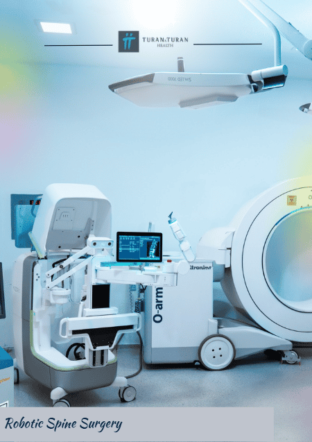
One of the persistent challenges in traditional neurosurgery is accounting for intraoperative anatomical shifts. When patient positioning changes or soft tissues undergo surgical modification, preoperative static scans may no longer reflect the immediate reality inside the body. To solve this problem, Asklepios Kliniken Hamburg utilizes an imaging-guided robotic system that brings high-resolution diagnostic imaging directly into the operating suite during active surgery.
At [00:01:36], the video introduces this robot-assisted mobile imaging device, which features a large field of view. Capable of performing rapid intraoperative 2D X-rays and full 3D volumetric CT-quality scans, this system allows the surgical team to capture updated anatomical images in seconds without moving the patient out of the sterile field. As noted at [00:01:43], these high-resolution visualizations provide immediate feedback regarding bone cuts, implant alignment, and tissue resection boundaries.
Key Benefits of Intraoperative Mobile 3D Imaging
- In-Suite Verification: Confirms precise hardware placement and tumor resection before the surgical incision is closed.
- Elimination of Revision Surgeries: Immediate intraoperative imaging prevents post-operative surprises and avoids secondary corrective procedures.
- Dynamic Map Updating: Automatically updates spatial registration data on navigation monitors to reflect real-time anatomical changes.
- Time and Workflow Efficiency: Saves valuable operating room time while granting the surgical team greater flexibility and control.
As highlighted at [00:01:49], this real-time verification capability saves crucial operating time while offering the surgical team significant operational freedom. Surgeons no longer need to wait until post-operative recovery to verify whether an implant is perfectly positioned. If a minor alignment adjustment is required, it can be identified and corrected immediately, virtually eliminating the need for costly and uncomfortable secondary revision surgeries.
Central Command: The Integrated Mobile Workstation Platform
Managing multiple advanced digital components requires a centralized hub to collect, synchronize, and display vital patient metrics. The entire Brainlab ecosystem at Asklepios Kliniken Hamburg is linked together through an advanced surgical education and operating room platform. As detailed at [00:01:56], this platform centers around a mobile workstation equipped with tracking cameras, a high-resolution 32-inch display, and a responsive 4K touchscreen visualization interface.
This centralized platform acts as the nerve center of the operating room, consolidating diagnostic imaging, real-time optical tracking feedback, endoscopic video feeds, and anesthesia monitoring into a single unified dashboard at [00:02:12]. Having all critical patient data aggregated onto a single high-definition screen streamlines communication across the surgical team, ensuring that neurosurgeons, scrub nurses, and anesthesiologists share identical situational awareness throughout every phase of the procedure.
| Hardware Component | Technical Specifications | Functional Role in the Operating Room |
|---|---|---|
| Mobile Workstation | High-mobility, ergonomic design with integrated camera mount | Houses tracking hardware and core processing units. |
| 4K Touchscreen Display | 32-inch high-resolution ultra-HD interface | Aggregates multi-source patient data, 3D plans, and live video. |
| Remote Monitoring Suite | Encrypted, high-bandwidth digital streaming integration | Enables real-time remote consultations and efficient documentation. |
Furthermore, the digital architecture of this workstation supports remote monitoring and efficient electronic documentation at [00:02:14]. Senior specialists and consulting surgeons can observe procedural progress securely from outside the immediate operating room, providing expert input when needed. Automatically logging surgical milestones and intraoperative imaging logs directly into the patient's electronic medical record further enhances clinical accountability and administrative efficiency.
Translating Technology to Care: Clinical Advantages for Brain and Spine Patients
While the technological capabilities of Brainlab robotic systems are impressive, their ultimate metric of success is measured in improved patient outcomes. Integrating computer-assisted navigation and robotic guidance into neurosurgery delivers concrete, transformative clinical benefits for patients undergoing brain and spinal treatments.
By replacing traditional, highly invasive open surgical procedures with minimally invasive robot-guided techniques, surgeons can achieve their treatment objectives through significantly smaller incisions. Sub-millimeter accuracy allows surgeons to avoid vital vascular structures and healthy neurological pathways, leading to reduced operative blood loss, lower post-operative pain levels, and a decreased risk of surgical site infections.
Direct Patient Benefits of Integrated Robotic Surgery
- Minimally Invasive Interventions: Smaller skin incisions result in minimal muscular disruption and less post-operative discomfort.
- Enhanced Neurological Protection: Precision guidance helps protect critical functional areas of the brain and spinal cord.
- Faster Recovery and Rehabilitation: Patients generally experience shorter hospital stays and a quicker return to daily routines.
- Minimized Radiation Exposure: Targeted intraoperative imaging protocols optimize diagnostic output while reducing unnecessary radiation exposure.
For patients facing complex neurosurgical diagnoses, seeking care at an institution equipped with advanced technology offers deep reassurance. The seamless combination of human expert judgment and state-of-the-art robotic precision ensures that every surgical step is planned, simulated, executed, and verified with extreme rigor, cementing the leadership of robotic surgery in Germany.
Leading the Future of Healthcare: Asklepios Kliniken Hamburg
The installation of the complete Brainlab surgical and robotics platform at Asklepios Kliniken Hamburg represents a landmark milestone in the evolution of modern medicine. As robotic platforms continue to incorporate machine learning algorithms, real-time tissue analytics, and augmented reality overlays, the line between human skill and technological augmentation will continue to yield safer, more effective surgical options.
By pioneering this nationwide integration, Hamburg has positioned itself at the global forefront of medical innovation. Patients from around the world look to medical centers like Asklepios Kliniken Hamburg for high-standard care in specialized neurosurgery, orthopedics, and complex spinal reconstruction. The success of this integrated operating suite demonstrates that when cutting-edge technology is deployed thoughtfully, it enhances the surgeon's skills and elevates the safety and well-being of every patient who enters the operating room.
As described at [00:02:19], the Brainlab family of robotic surgery solutions represents a true paradigm shift. By merging digital planning, 3D smart visualization, steady robotic arm assistance, dynamic intraoperative imaging, and centralized control into a unified surgical ecosystem, Asklepios Kliniken Hamburg continues to define the future of robotic surgery in Hamburg Germany and set international benchmarks for patient care.
Ready to Explore Robotic Surgery Options in Germany?
PlacidWay Medical Tourism connects you with premier hospitals and world-renowned surgical specialists for Robotic Surgery in Germany at Asklepios Kliniken Hamburg. Access advanced robotic surgery treatments tailored to your medical needs. Contact us today for personalized guidance.
GET A FREE QUOTEView Full Video Transcript
[00:00:00] Germany's first clinic to have all of these components from FDP, Skrynik, Schiller and Halsen for the first time nationwide, all the components of the comprehensive Brainlab surgical system and robotics are available in one clinic.
[00:00:14] It feels like being in a science fiction world, like Star Trek and the Enterprise. A navigation software system, digital devices, specialized robotic modules, and a complete package make neurosurgical procedures on the brain and spine even more precise, with an even greater focus on patient safety.
[00:00:31] Let's now take a closer look at these intelligent technologies and smart assistants.
[00:00:35] The core planning software enables better planning and better execution of procedures. For example, you can individually mark areas on a patient's 3D images, create 3D image animations, and precisely work through surgical cuts, almost like a simulator directly on the PC.
[00:00:53] The plan can then be transferred directly to the operating room. [Music]
[00:00:58] The system is complemented by 3D glasses. This software enables completely new perspectives for planning and anatomy.
[00:01:05] The patient's disease can be presented in 3D in a completely different way, making it much more visual and easier for patients to understand. It is also extremely useful for training and education.
[00:01:17] Here we have the robotic arm. Every task requires a high degree of fine motor skills, precision, and endurance. That is exactly what the robotic arm is designed for.
[00:01:27] It is modeled after the human arm, and it can be used, for example, to position implants much more precisely and in a more targeted manner, allowing the surgeon to focus entirely on the procedure.
[00:01:36] The imaging-guided robotic system combines a robot-assisted mobile imaging device with a large field of view.
[00:01:43] It enables high-resolution X-ray images and 2D and 3D visualizations during surgery. This saves time and gives the surgical team greater freedom while maintaining control.
[00:01:56] The system is complemented by a modern surgical education and surgical platform. It consists of a mobile workstation with a camera, a high-resolution 32-inch display, a 4K touchscreen visualization system, where all patient data comes together.
[00:02:14] The system can be monitored from anywhere, allowing checks and documentation to be carried out efficiently.
[00:02:19] This is the Brainlab family of robotic surgery solutions. [Music]























Share this listing